
Figures & data
Figure 1. The abundance of F. nucleatum is associated with the progression and prognosis of CRC. (A) The abundance of F. nucleatum was measured in 30 pairs of CRC tissues. (B) Heatmap illustrating the association of various histopathological features and high-abundance and low-abundance F. nucleatum tumors. (C) Recurrence-free survival (RFS) was compared between groups with low and high F. nucleatum abundance. (D) Receiver operating characteristic (ROC) analysis was performed according to F. nucleatum abundance and AJCC in CRC.
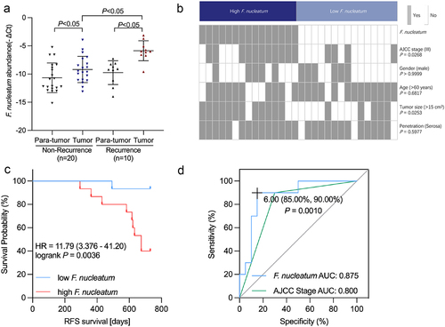
Figure 2. F. nucleatum represses chemotherapy-induced pyroptosis in CRC. (a) Representative bright-field images of CRC cells cocultured with chemotherapy drugs and F. nucleatum. Yellow arrows indicate large bubbles emerging from the cell membrane. (b) Transmission electron microscopy observation of CRC cells cocultured with 5-fu and F. nucleatum. Red and orange arrows indicate cell membrane pores and F. nucleatum, respectively. (c) The effects of chemotherapy drugs and F. nucleatum on LDH release from cells were detected. (d-e) the effect of F. nucleatum and chemotherapy on the expression of pyroptosis-related proteins in CRC cells was detected by Western blot. (f) Cell viability after treatment with chemotherapy and F. nucleatum in CRC cells was detected by CCK8 assay. All experiments were performed in biological triplicates.
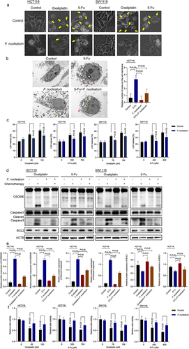
Figure 3. BCL2 knockdown affects the inhibitory effect of F. nucleatum on chemotherapy-induced pyroptosis. (a-b) the effect of F. nucleatum on the expression of pyroptosis-related proteins in chemotherapy-treated CRC cells after inhibition of BCL2 expression was detected by Western blotting. (c) The morphological changes in F. nucleatum-treated CRC cells after BCL2 inhibition were observed under a light microscope. (d) LDH release from chemotherapy-treated CRC cells after F. nucleatum intervention after BCL2 inhibition was detected. All experiments were performed in biological triplicates.
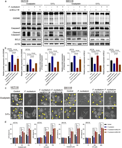
Figure 4. F. nucleatum induces chemotherapy resistance by regulating BCL2. (a) Survival of chemotherapy-treated CRC cells after F. nucleatum intervention and BCL2 inhibition was determined by CCK8 assay. (b) Representative images of CRC patient-derived organoids with F. nucleatum or BCL2 shRNA. (c) The size of proliferation on CRC patient-derived organoids was measured. (d) LDH release from CRC patient-derived organoids with F. nucleatum after BCL2 downregulation was detected. (e) Xenograft tumors in the nude mouse model were used to examine the effect of BCL2 inhibition on the chemoresistance of F. nucleatum-treated CRC cells. (f) Tumor volumes were calculated after injection every 3 days. (g) Tumor weights are represented as the means of tumor weights ± S.D. (h) The release of serum LDH in nude mice was measured. Bars indicate S.D.
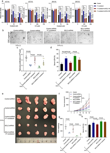
Figure 5. F. nucleatum affects BCL2-mediated pyroptosis by regulating YAP. (a) ChIP assay to detect the enrichment of YAP in the BCL2 promoter region in chemotherapeutic drug-treated CRC cells after F. nucleatum intervention. (b-c) CRC cells were transfected with si-YAP. After coculturing with F. nucleatum and chemotherapy drugs, pyroptosis target proteins were detected by western blotting. (d) Representative bright-field images were observed under a light microscope. Yellow arrows indicate large bubbles emerging from the cell membrane. (e) The effect of YAP knockdown on LDH release in CRC cells infected with F. nucleatum was detected. All experiments were performed in biological triplicates.
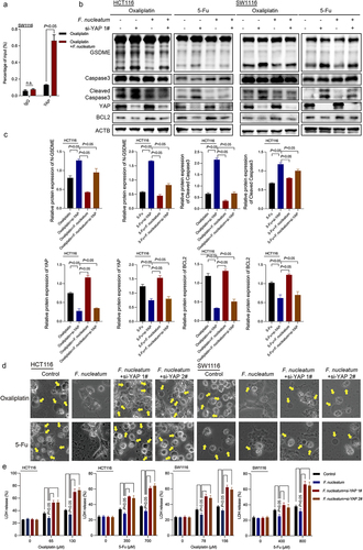
Figure 6. YAP mediates F. nucleatum-induced chemoresistance. (a) CCK8 assay was used to detect the effect of si-YAP on the viability of CRC cells treated with chemotherapy drugs after F. nucleatum intervention. (b) Representative images of chemotherapy-treated CRC patient-derived organoids with F. nucleatum, YAP shRNA or BCL2 adenovirus. (c) The size of proliferation on chemotherapy-treated CRC cells CRC patient-derived organoids was measured. (d) LDH release from CRC patient-derived organoids with F. nucleatum after YAP downregulation or BCL2 upregulation was detected. (e) Representative data of xenograft tumors in the nude mouse model bearing HCT116 cells in different groups. (f-g) statistical analysis of mouse tumor volumes (f) and weights (g). (h) The release of LDH in the serum of nude mice was detected. Bars indicate S.D.
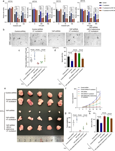
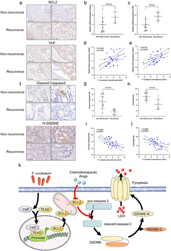
Figure 7. F. nucleatum, YAP, BCL2, caspase-3 and GSDME were clinically relevant. (a) Protein levels of YAP and BCL2 in CRC tissues from non-recurrent and recurrent patients were detected by immunohistochemistry. (b and c) the mRNA expression of YAP and BCL2 was statistically analyzed by qRT‒PCR. (d and e) correlation of the expression levels of F. nucleatum, YAP and BCL2 in human CRC tissues. (f) Protein levels of cleaved caspase-3 and N-GSDME in CRC tissues from non-recurrent and recurrent patients were detected by immunohistochemistry. (g and h) statistical analysis of immunohistochemical immunoreactive score of Remmele and Stegner (IRS) scores of cleaved caspase-3 and N-GSDME proteins. (i and j) correlation of the expression levels of F. nucleatum, cleaved caspase-3 and N-GSDME in human CRC tissues. (k) The schematic model shows that F. nucleatum may affect chemoresistance in CRC cells by inhibiting chemotherapy-induced pyroptosis.
Supplementary Figure S2.tif
Download TIFF Image (9.7 MB)Supplementary table S1 The list of primers and siRNA sequence.xlsx
Download MS Excel (10.2 KB)Supplementary Figure Legends clean.docx
Download MS Word (21.8 KB)Supplementary Figure S5.tif
Download TIFF Image (7.3 MB)Supplementary Figure S1.tif
Download TIFF Image (13.5 MB)Supplementary Figure S3.tif
Download TIFF Image (5.3 MB)Supplementary Figure S4.tif
Download TIFF Image (9.8 MB)Supplementary Figure S6.tif
Download TIFF Image (4.7 MB)Data availability statement
Data supporting these findings are available from the corresponding author upon reasonable request.