Dietary restriction rescues 5-fluorouracil-induced lethal intestinal toxicity in old mice by blocking translocation of opportunistic pathogens
Duozhuang Tanga Jiangxi Key Laboratory of Clinical and Translational Cancer Research, Department of Oncology, The Second Affiliated Hospital of Nanchang University, Nanchang, Jiangxi, China;b Department of Hematology, The Second Affiliated Hospital of Nanchang University, Nanchang, Jiangxi, ChinaView further author information
,
Rongrong Qiua Jiangxi Key Laboratory of Clinical and Translational Cancer Research, Department of Oncology, The Second Affiliated Hospital of Nanchang University, Nanchang, Jiangxi, ChinaView further author information
,
Xingxing Qiua Jiangxi Key Laboratory of Clinical and Translational Cancer Research, Department of Oncology, The Second Affiliated Hospital of Nanchang University, Nanchang, Jiangxi, ChinaView further author information
,
Man Suna Jiangxi Key Laboratory of Clinical and Translational Cancer Research, Department of Oncology, The Second Affiliated Hospital of Nanchang University, Nanchang, Jiangxi, ChinaView further author information
,
Mingyue Sua Jiangxi Key Laboratory of Clinical and Translational Cancer Research, Department of Oncology, The Second Affiliated Hospital of Nanchang University, Nanchang, Jiangxi, ChinaView further author information
,
Zhendong Taoc Department of Medical Laboratory Medicine, Jiangxi Province Hospital of Integrated Chinese & Western Medicine, Nanchang, Jiangxi, ChinaView further author information
Si Taoa Jiangxi Key Laboratory of Clinical and Translational Cancer Research, Department of Oncology, The Second Affiliated Hospital of Nanchang University, Nanchang, Jiangxi, ChinaCorrespondence[email protected] View further author information
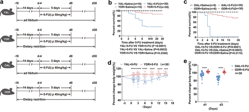
Figure 1. DR increases survival of 5-FU treated mice.
(A) Scheme of experiments. Young mice (2-month-old) and old mice (20–24-month-old) were exposed to AL diet or DR for 14 days before intraperitoneal 5-FU injection which was daily performed for 5 days, and the diet regimen was continued afterward. For the control group, saline was injected instead of 5-FU.
(B,C) Survival was monitored daily after 5-FU treatment (Data combined from 3 independent experiments. n = 30 mice per group for the AL + 5-FU group and DR + 5-FU group; n = 10 mice per group for the young AL+Saline group and young DR+Saline group; n = 6 mice per group for the old AL+Saline group and old DR+Saline group).
(D,E) Percent change of body weight at indicated timepoints after 5-FU treatment compared to before 5-FU treatment (n = 30 mice per group. Data combined from 3 independent experiments).
(B,C) Gehan-Breslow-Wilcoxon test; (D,E) Unpaired two-tailed, Student’s t test. Results were displayed as mean±SD. **p < .01; **** p < .0001. YAL+Saline: young mice on AL diet and received saline injection; YDR+Saline: young mice on DR diet and received saline injection; YAL + 5-FU: young mice on AL diet and received 5-FU injection; YDR + 5-FU: young mice on DR diet and received 5-FU injection. OAL+Saline: old mice on AL diet and received saline injection; ODR+Saline: old mice on DR diet and received saline injection; OAL + 5-FU: old mice on AL diet and received 5-FU injection; ODR + 5-FU: old mice on DR diet and received 5-FU injection; d1: day 1 after 5-FU injection; d3: day 3 after 5-FU injection.
Figure 2. DR inhibits 5-FU-induced intestinal inflammation.
Young mice (2-month-old) and old mice (20–24-month-old) were exposed to AL diet or DR for 14 days before intraperitoneal 5-FU injection which was daily performed for 5 days (day -4–day 0), and the diet regimen was continued afterward. For the control group, saline was injected instead of 5-FU. Mice were sacrificed at certain timepoints and small intestinal tissue was collected for further analysis.
(A,B) Representative photos of mice from indicated groups. Note that the intestines of AL mice exposed to 5-FU were covered with mucus and exhibited significant edema, multiple erosions, and hardly visible formed stools, while the intestines of DR mice treated with 5-FU looked similar to saline treated control mice with no visible damage in the intestinal wall and lots of formed stools (arrows).
(C,D) Bowen scoring system determined diarrhea score indexes from indicated groups (n = 30 mice per group. Data combined from three independent experiments).
(E-J) Relative expression of indicated genes in duodenum, jejunum, and ileum tissues collected on day 3 after 5-FU treatment by qRT-PCR (n = 5 mice per group randomly picked from 2 independent experiments).
(C,D) Unpaired two-tailed, Student’s t test; (E-J) One-way ANOVA test. Results were displayed as mean±SD. *p < .05; **p < .01;***p < .001; ****p < .0001; ns, not significant.
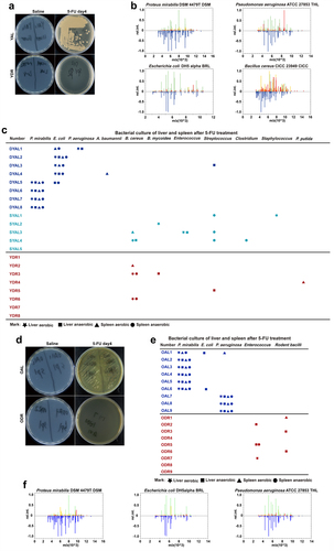
Figure 3. DR inhibits intestinal opportunistic pathogens translocation after 5-FU treatment.
Young mice and old mice (20–24-month-old) were exposed to AL diet or DR for 14 days before intraperitoneal 5-FU injection which was daily performed for 5 days (day -4–day 0), and the diet regimen was continued afterward. For the control group, saline was injected instead of 5-FU. Mice were sacrificed on day 4 after 5-FU treatment and liver and spleen were collected and homogenized for bacterial culture and further analysis.
(A,D)
Representative pictures of bacteria culture dishes from indicated groups.
(B,F) The bacterial colonies grown out from the liver and spleen homogenates were identified by mass spectrometer. Above the ordinate 0 scale is the key mass spectral peak data of the target bacteria, which is compared with the known strain spectrum in the database located below the ordinate 0 scale identifying specific bacteria.
(C,E) Bacterial species identified from colonies grown out from individual mouse tissue homogenates. (Data combined from two independent experiments). Mice which met Death or Moribundity Criteria according to Guidelines for Endpoints in Animal Study Proposals after 5-FU were defined as non-survived or dead. Mice which did not meet Death or Moribundity Criteria were defined as survived. DYAL: dead young ad libitum; SYAL: survived young ad libitum.
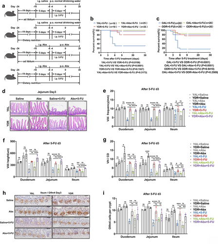
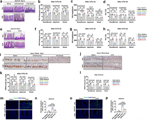
Figure 4. Abx administration rescued survival of AL mice after 5-FU treatment.
(A) Scheme of experiments. Young mice (2-month-old) and old mice (20–24-month-old) were exposed to AL diet or DR for 14 days before intraperitoneal 5-FU injection which was daily performed for 5 days (day-4–day0), and the diet regimen was continued afterward. Seven days (day-10) prior to 5-FU administration, mice were given a gavage of broad-spectrum antibiotics (Abx group) or saline (control group) for 5 days and then in drinking water for the following days.
(B,C) Survival was monitored daily after 5-FU treatment (Data combined from 2 independent experiments. n = 10 mice per group for the young AL + 5-FU group and DR + 5-FU group; n = 25 mice per group for the young AL+Abx +5-FU group and DR+Abx +5-FU group; n = 20 mice per group for the old AL + 5-FU group, DR + 5-FU group, AL+Abx +5-FU group, and DR+Abx +5-FU group).
(D) Representative images of H&E staining of jejunum on day 3 after 5-FU treatment from indicated groups. Scale bar: 50 μm.
(E-G) Villi frequencies, villi height, and VH/CD (the ratio of villi height versus crypt depth) (n = 5 mice per group randomly picked from 2 independent experiments).
(H) Representative images of Immunohistochemistry staining of Olfm4 of ileum on day 3 after 5-FU treatment from indicated groups. Scale bar: 20 μm.
(I) Basal crypt Olfm4-positive cell number per crypt on day 3 after 5-FU treatment
(n = 5 mice per group randomly picked from 2 independent experiments).
(B,C) Gehan-Breslow-Wilcoxon test; (E-G, I) One-way ANOVA test. Results were displayed as mean±SD. *p < .05; **p < .01;***p < .001; ****p < 0.0001; ns, not significant. YAL+Saline: young mice on AL diet and received saline injection; YDR+Saline: young mice on DR diet and received saline injection; YAL + 5-FU: young mice on AL diet and received 5-FU injection; YDR + 5-FU: young mice on DR diet and received 5-FU injection;YAL+Abx +5-FU: young mice on AL diet and received a gavage of Abx and 5-FU injection; YDR+Abx +5-FU: young mice on DR diet and received a gavage of Abx and 5-FU injection. d3: day3 after 5-FU injection; Abx: broad-spectrum antibiotics.
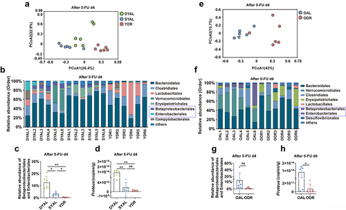
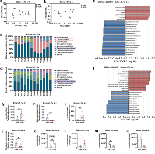
Figure 5. DR reduced the opportunistic pathogens in the gut microbiota of mice after 5-FU treatment.
Young mice (2-month-old) and old mice (20–24-month-old) were exposed to AL diet or DR for 14 days before intraperitoneal 5-FU injection which was daily performed for 5 days (day -4–day 0), and the diet regimen was continued afterward. Fecal samples were randomly collected from mice on day 4 after 5-FU injection for 16S rRNA gene sequencing (n = 6 mice per group randomly picked from 2 independent experiments) and qPCR analysis (n = 5 mice per group randomly picked from 2 independent experiments).
(A,E) Variation of intestinal microbiota structure of indicated groups along PC1 and PC2 of PCoA based on the Bray – Curtis distance.
(B,F) Relative abundance of the intestinal microbiota of indicated groups on the order level showed by 16S rRNA gene sequencing.
(C,J) Relative abundance of the Betaproteobacterales and Enterobacteriales showed by 16S rRNA gene sequencing.
(D,H) qPCR analysis of the amounts of Proteus of indicated groups.
(C,D) One-way ANOVA test;(G,H) Unpaired two-tailed,Student’s t test; Results were displayed as mean±SD; ns, not significant; * p < .05; **p < 01. Mice which met Death or Moribundity Criteria according to Guidelines for Endpoints in Animal Study Proposals after 5-FU were defined as non-survived or dead. Mice which did not met Death or Moribundity Criteria were defined as survived. DYAL: dead young ad libitum; SYAL: survived young ad libitum.
Figure 6. DR protects intestinal physical barrier from 5-FU.
Young mice (2-month-old) and old mice (20–24-month-old) were exposed to AL diet or DR for 14 days before intraperitoneal 5-FU injection which was daily performed for 5 days (day -4–day 0), and the diet regimen was continued afterward. For the control group, saline was injected instead of 5-FU. The small intestines were collected on day3 after 5-FU treatment for further analysis (n = 5 mice per group randomly picked from 2 independent experiments).
(A,E) Representative images of H&E straining of ileum from indicated groups. Scale bar: 50 μm.
(B-H) Villi frequencies, villi height, and VH/CD (the ratio of villi height versus crypt depth) (n = 5 mice per group randomly picked from 2 independent experiments).
(I,J) Representative images of Immunohistochemistry staining of Olfm4 of ileum. Scale bar: 20 μm.
(K,L) Basal crypt Olfm4-positive cell number per crypt in indicated fractions of the intestine from indicated groups (30 crypts were counted for each fraction. n = 5 mice per group randomly picked from 2 independent experiments).
(M,O) Representative images of TUNEL staining of jejunum. Scale bar: 20 μm.
(N,P) Crypt apoptotic cell number per crypt in jejunum as determined by TUNEL staining.
(B-D,F-H,K,L,N,P) One-way ANOVA test. Results were displayed as mean±SD. *p < .05; **p < .01;***p < .001; ****p < .0001; ns, not significant. Mice which met Death or Moribundity Criteria according to Guidelines for Endpoints in Animal Study Proposals after 5-FU were defined as non-survived or dead. Mice which did not met Death or Moribundity Criteria were defined as survived. DYAL: dead young ad libitum; SYAL: survived young ad libitum.
Figure 7. DR protects intestinal biological barrier from 5-FU.
Fecal samples were randomly collected from in young and old mice after two weeks of AL or DR diet before 5-FU treatment and gut microbiota were analyzed by 16S rRNA gene sequencing (n = 6 mice per group randomly picked from 2 independent experiments) and qPCR analysis (n = 5 mice per group randomly picked from 2 independent experiments).
(A,B) Principal-coordinate analysis (PCoA) based on Bray-Curtis distance showed a significant shift of the overall structure of the gut microbiota by DR in both young and old mice.
(C,D) Relative abundance of the intestinal microbiota of indicated groups on the order level showed by 16S rRNA gene sequencing.
(E,F) LDA scores in the fecal microbiomes of indicated groups. LDA score > 4 and top fifteen bacteria were shown.
(G-J) Relative abundance of the Lactobacillales and Betaproteobacteriales showed by 16S rRNA gene sequencing.
(K-N) qPCR analysis of the amounts of Lactobacillus and Proteus of indicated groups.
(G-N) Unpaired two-tailed, Student’s t test. Results were displayed as mean±SD. *p < .05; **p < .01;***p < .001; ns, not significant.
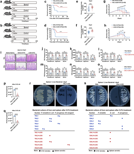
Figure 8. LGG gavage partially rescues survival of AL mice exposed to 5-FU.
(A,B) Scheme of experiments. Young mice (2-month-old) and old mice (20–24-month-old) were treated with LGG or saline by gastric gavage for 3 weeks before 5-FU treatment (1 × 10⁹ CFU LGG per mouse per day). Then intraperitoneal 5-FU or saline (control) injection was daily performed for 5 days(day -4,day 0). In the young group: data were combined from 3 independent experiments (n = 20 mice per group for the AL + 5-FU group, n = 30 mice per group for the AL+LGG +5-FU group). In the old group: data were combined from 3 independent experiments. (n = 25 mice per group for the AL + 5-FU group and the AL+LGG +5-FU group).
(C,D) Survival was monitored daily after 5-FU treatment.
(E,F) Percent change of bodyweight on day 3 after 5-FU treatment.
(G,H) Bowen scoring system determined diarrhea score indexes from indicated groups.
(I-O) Small intestines were collected for further analysis. (I) Representative images of H&E straining of Jejunum on day 3 after 5-FU treatment. Scale bar: 50 μm. (J-O) Villi frequencies, villi height, and VH/CD (the ratio of villi height versus crypt depth) (n = 5 mice per
group randomly picked from 2 independent experiments).
(P,Q) Mice were sacrificed on day 4 after 5-FU treatment and liver and spleen were collected and homogenized for bacterial culture and further analysis. Representative pictures of bacteria culture dishes from indicated groups were shown. The bacterial colonies grown out from the liver and spleen homogenates were identified by mass spectrometer. The bellowing tables show bacterial species identified from colonies grown out from individual mouse tissue homogenates (n = 6 mice per group from 2 independent experiments).
(R,S) Fecal samples were randomly collected on day 4 after 5-FU treatment. The amounts of Proteus were determined by qPCR analysis of (n = 6 mice per group from 2 independent experiments).
(C,D) Gehan-Breslow-Wilcoxon test; (E-H,P,Q) Unpaired two-tailed, Student’s t test; (J-O) One-way ANOVA test. Results were displayed as mean±SD. *p < .05; **p < .01;***p < .001; ****p < 0.0001; ns, not significant.
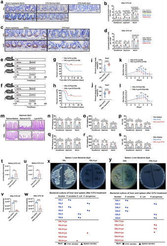
Figure 9. DR protects intestinal chemical barrier from 5-FU and lysozyme gavage partially rescues survival of AL mice exposed to 5-FU.
(A-D)Young mice (2-month-old) and old mice (20–24-month-old) were exposed to AL diet or DR for 14 days before intraperitoneal 5-FU injection which was daily performed for 5 days, and the diet regimen was continued afterward. For the control group, saline was injected instead of 5-FU. Small intestinal tissue were collected on day 3 after 5-FU treatment for immunohistochemistry staining of
Lysozyme. (A,C) Representative images of ileum. Scale bar: 20 μm. (B,D) Statistic analysis of number of Lysozyme-positive cells per crypt in indicated fractions of indicated groups (n = 5 mice per group randomly picked from 2 independent experiments).
(E,F) Scheme of experiments. (E) Young mice (2-month-old) and (F) old mice (20–24-month-old) were treated with lysozyme by gastric gavage for 2 weeks before 5-FU treatment. Then, intraperitoneal 5-FU or saline (control) injection was daily performed for 5 days (day -4,day 0). In the young group: data were combined from 3 independent experiments (n = 24 mice per group for the AL + 5-FU group and AL+Lyz +5-FU group). In the old group: data were combined from 3 independent expriments (n = 25 mice per group for the AL + 5-FU group and AL+Lyz +5-FU group). And the intestines were collected for further analysis (n = 5 mice per group randomly picked from 2 independent experiments).
(G,H) Survival was monitored daily after 5-FU treatment.
(I,J) Percent change of body weight on day 3 after 5-FU treatment.
(K,L) Bowen scoring system determined diarrhea score indexes from indicated groups.
(M-S) Small intestines were collected for further analysis. (M) Representative images of H&E straining of Jejunum on day 3 after 5-FU treatment. Scale bar: 50 μm. (N-S) Villi frequencies, villi height, and VH/CD (the ratio of villi height versus crypt depth) (n = 5 mice per group randomly picked from 2 independent experiments).
(P,Q) Mice were sacrificed on day4 after 5-FU treatment and liver and spleen were collected and homogenized for bacterial culture and further analysis. Representative pictures of bacteria culture dishes from indicated groups were shown. The bacterial colonies grown out from the liver and spleen homogenates were identified by mass spectrometer. The bellowing tables show bacterial species identified from colonies grown out from individual mouse tissue homogenates (n = 6 mice per group from 2 independent experiments).
(T-W) Fecal samples were randomly collected before (day -4) and after (day 4) 5-FU treatment. The amounts of Lactobacillus and Proteus were determined by qPCR analysis of (n = 6 mice per group from 2 independent experiments).
(G,H) Gehan-Breslow-Wilcoxon test; (I-L,T-W) Unpaired two-tailed, Student’s t test; (B,D,N-S) One-way ANOVA test. Results were displayed as mean±SD. *p < .05; **p < .01;***p < .001; ****p < .0001; ns, not significant.
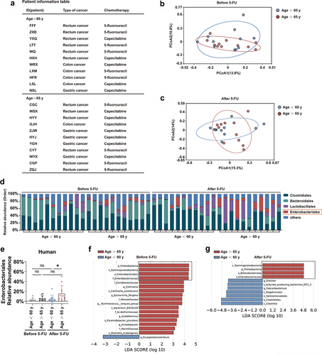
Figure 10. Relative abundance of opportunistic pathogens increases after 5-FU chemotherapy in old patients.
Fecal samples of patients with gastrointestinal cancers were collected for 16S rRNA gene sequencing before the first cycle of 5-FU chemotherapy and one week/two weeks after the 5-FU treatment (n = 11 patients in the group aging less than 60 years old, and n = 11 patients in the group aging over 65 years old).
Patient information.
(B,C) Variation of intestinal microbiota structure of indicated groups along PC1 and PC2 of PCoA based on the Bray – Curtis distance.
(D) Relative abundance of the intestinal microbiota of indicated groups on the order level showed by 16S rRNA gene sequencing.
(E) Relative abundance of the Enterobacteriales showed by 16S rRNA gene sequencing.
(F,G) LDA scores in the fecal microbiomes of indicated groups. LDA score > 3 and top seventeen bacteria were shown.
(E) One-way ANOVA test, Results were displayed as mean±SD; *p < .05; ns, not significant.