
Figures & data
Figure 1. RstAB promotes LF82 virulence in chronic colitis mice.
(a) DSS induced mouse model of chronic colitis.
(b-e) Bacterial titers of LF82 in mouse colon (b), cecum (c), ileum (d) and jejunum (e) infected with WT, ΔrstA, ΔrstAB, WT+pWSK129, ΔrstA+pWSK129, ΔrstAB+pWSK129, ΔrstA+prstA and ΔrstAB+prstAB at 24 h p.i. (n = 10).
Horizontal bars indicate the median. Data were obtained from three independent experiments and analyzed using the two-sided Mann – Whitney U test to calculate P values. *P< .05, **P < .01, *** P < .001; n.s., not significant.
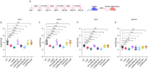
Figure 2. RstAB promotes the replication of LF82 in macrophages.
(a-b) qRT-PCR analyses the genes expression levels of rstA (a) and rstB (b) in LF82 cultured in RPMI 1640 medium or infected Raw 264.7 cells for 1, 6 and 24 h.
(c) Bacterial titers of WT, ΔrstA, ΔrstAB, WT+pWSK129, ΔrstA+pWSK129, ΔrstAB+pWSK129, ΔrstA-prstA or ΔrstAB-prstAB infected Raw 264.7 cells for 6 or 24 h relative to 1 h.
(d) Confocal microscopy determined the bacterial number of WT, ΔrstA, ΔrstAB, WT+pWSK129, ΔrstA+pWSK129, ΔrstAB+pWSK129, ΔrstA-prstA or ΔrstAB-prstAB in Raw 264.7 cells at 1, 6 or 24 h p.i.
(e) Bacterial titers of WT, ΔrstA, ΔrstAB, WT+pWSK129, ΔrstA+pWSK129, ΔrstAB+pWSK129, ΔrstA-prstA and ΔrstAB-prstAB infected MBMM for 6 or 24 h relative to 1 h.
The number of bacteria at 6 and 24 h p.i. was compared with that at 1 h p.i. by LB solid plate counting. The replication multiples at 6 and 24 h p.i. were expressed by T6/T1 and T24/T1, respectively.
Data were obtained from three independent experiments and analyzed using Student’s t-test. *P< .05; **P < .01; ***P < .001; n.s., not significant.

Figure 3. RstAB promotes LF82’s replication in macrophages by regulating the expression of csgD and asr.
(a) COG analysis of DEGs in ΔrstAB when infected Raw 264.7 cells compared with that of WT at 1 h p.i.
(b) qRT-PCR analysis of the gene expression of DEGs in ΔrstAB when infected Raw 264.7 cells compared with that of WT at 1 h p.i.
(c) asr gene expression of WT and ΔrstAB when infected Raw 264.7 cells at 1 h p.i.
(d) csgD gene expression of WT and ΔrstAB when infected Raw 264.7 cells at 1 h p.i.
(e) Heatmap of asr and csgABCDEFG genes in ΔrstAB and WT when infected Raw 264.7 cells at1 h p.i.
(f) Volcano plot showing DEGs in the transcriptome of ΔrstAB when infected Raw 264.7 cells compared with that of WT at 1 h p.i.
Data were obtained from three independent experiments and analyzed using Student’s t-test. *P < .05, **P < .01, ***P < .001; n.s., not significant.
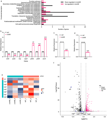
Figure 4. RstA regulates biofilm formation of LF82 by directly activates the expression of csgD.
(a-b) EMSAs of the binding of the promoter of csgD DNA fragment with purified RstA protein (a). rpoS promotor was used as the negative control (b). Images are representative of three independent experiments.
(c-d) EMSAs of the binding of purified RstA protein to the csgD promoter without RstA-box (c) or scrambled mutant of RstA-box (d). Images are representative of three independent experiments.
(e) Bacterial titers of WT, ΔcsgD, ΔrstABΔcsgD and ΔrstAB+pcsgD in Raw 264.7 cells at 6 or 24 h relative to 1 h p.i. The replication multiples at 6 and 24 h p.i. were expressed by T6/T1 and T24/T1, respectively.
(f) Confocal microscopy determined bacterial number of WT, ΔcsgD, ΔrstABΔcsgD and ΔrstAB+pcsgD in Raw 264.7 cells at 1, 6 or 24 h p.i.
(g) Bacterial titers of WT, ΔcsgD, ΔrstABΔcsgD and ΔrstAB+pcsgD in MBMM at 6 or 24 h relative to 1 h p.i. The replication multiples at 6 and 24 h p.i. were expressed by T6/T1 and T24/T1, respectively.
(h) Biofilm formation of WT, ΔrstA, ΔrstAB, ΔcsgD, ΔrstABΔcsgD and ΔrstAB+pcsgD in LB and RPMI 1640 medium.
Data were obtained from three independent experiments and analyzed using Student’s t-test. *P < .05, **P< .01, ***P < .001; n.s., not significant.
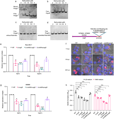
Figure 5. RstAB regulates the acid stress response of LF82 by directly activates the expression of asr.
(a-b) EMSAs of the binding of the promoter of asr DNA fragment with purified RstA protein (a). rpoS promotor was used as the negative control (b). Images are representative of three independent experiments.
(c-d) EMSAs of the binding of purified RstA protein to the asr promoter without RstA-box (c) or scrambled mutant of RstA-box (d). Images are representative of three independent experiments.
(e) Bacterial titers of WT, Δasr, ΔrstABΔasr and ΔrstAB+pasr in Raw 264.7 cells at 6 or 24 h relative to 1 h p.i. The replication multiples at 6 and 24 h p.i. were expressed by T6/T1 and T24/T1, respectively.
(f) Confocal microscopy determined bacterial number of WT, Δasr, ΔrstABΔasr and ΔrstAB+pasr in Raw 264.7 cells at 1, 6 or 24 h p.i.
(g) Bacterial titers of WT, Δasr, ΔrstABΔasr and ΔrstAB+pasr in MBMM at 6 or 24 h relative to 1 h p.i. The replication multiples at 6 and 24 h p.i. were expressed by T6/T1 and T24/T1, respectively.
(h) The tolerance of WT, ΔrstA, ΔrstAB, Δasr, ΔrstABΔasr and ΔrstAB+pasr to low pH, determined by count of viable bacteria at indicated time points.
Data were obtained from three independent experiments and analyzed using Student’s t-test. *P < .05, **P < .01, ***P < .001; n.s., not significant.
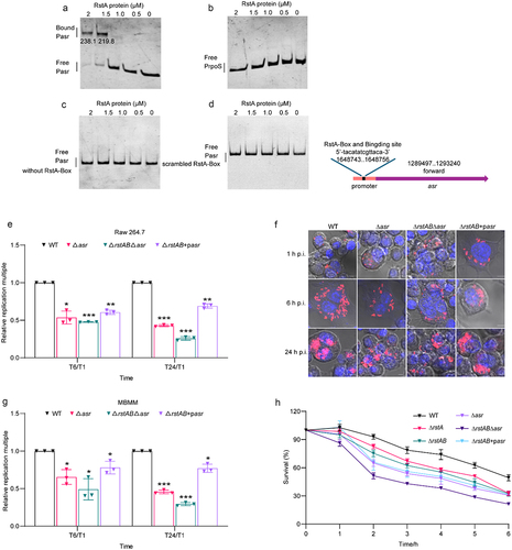
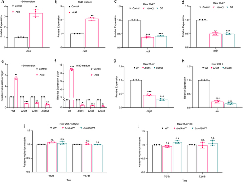
Figure 6. RstAB responds to acidic signal in macrophages and promotes intracellular replication of LF82.
(a-b) qRT-PCR analyses the gene expression levels of rstA (a) and rstB (b) in WT cultured in acidic RPMI 1640 medium relative to neutral RPMI 1640 medium.
(c-d) qRT-PCR analyses the genes expression of rstA (c) and rstB (d) in WT infected Raw 264.7 cells treatment with NH4Cl or CQ relative to treatment without NH4Cl and CQ at 1 h p.i.
(e-f) qRT-PCR analyses the gene expression levels of asr (e) and csgD (f) in WT, ΔrstA, ΔrstB and ΔrstAB cultured in acidic RPMI 1640 medium relative to neutral RPMI 1640 medium.
(g-h) qRT-PCR analyses the genes expression of asr (g) and csgD (h) in ΔrstA or ΔrstAB relative to WT infected Raw 264.7 cells at 1 h p.i.
(i-j) Bacterial titers of WT, ΔrstA or ΔrstAB in Raw 264.7 cells treatment with NH4Cl (i) or CQ (j) relative to treatment without NH4Cl or CQ at 6 or 24 h relative to 1 h p.i.
The number of viable bacteria at 6 and 24 h p.i. was compared with that at 1 h p.i. The replication multiples at 6 and 24 h p.i. were expressed by T6/T1 and T24/T1, respectively.
Data were obtained from three independent experiments and analyzed using Student’s t-test. *P < .05, **P < .01, ***P < .001; n.s., not significant.
Supplemental material
Supplemental Material
Download Zip (391.1 KB)Data availability statement
RNA sequencing data generated in this study are available from the NCBI SRA database. Accession to cite SRA data: PRJNA984413 https://www.ncbi.nlm.nih.gov/bioproject/?term=PRJNA984413.