Figures & data
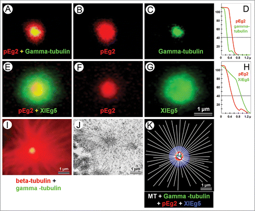
FIGURE 1. Localization of different proteins in prophase centrosome area in XL2 cells. (A–C) pEg2/gamma-tubulin double immunofluorescent labeling; (D) the profiles of the fluorescence intensity of pEg2 and gamma-tubulin immunofluorescence (vertical axis, arbitrary units) as a function of the distance from the geometric center of the fluorescent area (horizontal axis, µm); (E–G) pEg2/XlEg5 double immunofluorescent labeling; (H) the profile of the fluorescence intensity of pEg2 and XlEg5 immunofluorescence (vertical axis, arbitrary units) as a function of distance from the geometric center of the fluorescent area (horizontal axis, µm); (I) double β-tubulin/gamma-tubulin immunofluorescent labeling; (J) ultrastructure of centrosome region in prophase XL2 cell, TEM analysis; (K) representative scheme of different centrosomal proteins localization. Horizontal dashed line (D, H) shows the fluorescence level for which the measurement of the fluorescent spot radius was made. Scale bar 1 µm.