
Figures & data
FIGURE 1. The cell itself and the ECM it generates modulate the cell's mechanical milieu at multiple length scales. (A) Transmission electron microscope image of an osteocyte process traversing the plane of the image and orthogonal to the plane in the right upper image half, superimposed with computational fluid dynamics predictions of pericellular flow at cell surfaces. In terminally differentiated osteocytes, the cell processes and local ECM amplify the transduction of mechanical cues via pericellular fluid flow. Color plot represents flow field, where v is the flow velocity. Velocity (m/s) increases at sites where ECM ingresses into the pericellular space. Used with permission.Citation10 (B) Similar effects are observed around live model embryonic mesenchymal stem cells (C3H10T1/2, green) where flow fields are tracked using fluorescent microspheres (red). Used with permission.Citation11
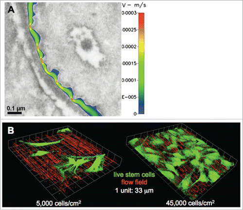
FIGURE 2. Relationship between motile and adherent stem cell states, MET/EMTs, and cell specialization through differentiation. The different states of the cell are depicted by a “cellular traffic light,” where motile states are depicted in green, adherent states in red, and transitional states in yellow. (A) Stem cells in adherent states may go through an MET or EMT to become motile. Osteocyte schematic used with permission.Citation27 (B) Once motile, circulating stem cells may again go through an MET or MET to become adherent.
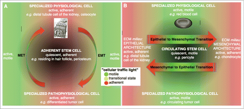
FIGURE 3. Retrospective mapping of the stem cells' mechanome, based on experimental data using an embryonic model mesenchymal stem cell line (C3H10T1/2). Actual stress and strain data points from experiments are represented as 95% confidence intervals, and color of resulting areas depicts the range of mechanical cues which correlating to early genetic markers of lineage commitment for chondrogenesis (yellow), chondrogenesis and haematopoesis (purple) and chondrogenesis, haematopoesis and osteogenesis (pink). In that sense, it maps states statistically conducive to fate. The next important step is to test prospectively whether those regions can be used to map libraries of mechanical cues to guide fate. Used with permission.Citation23
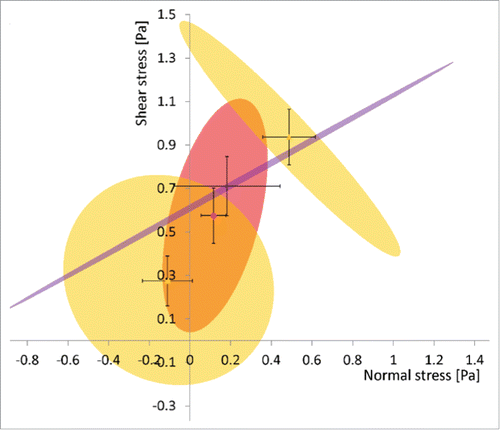
FIGURE 4. Experimental approach of Zile et al. to measure constitutive properties, including elastic properties (A-C) and viscous damping (D-F) of isolated cardiac muscle cells, in relation to cytoskeletal adaptation, i.e. microtubule and myofilament activation, in healthy and pressure overload hypertrophied hearts. (A-C) First the authors investigate passive spring properties of the 2 population of cells, by minimizing myofilament activation through chemical treatment while applying force to the gel in which cells are embedded. (D) The energy expended is then compared with that returned (E) to estimate viscous damping (F). Work refers to the process of applying a force over a distance; energy is the ‘cost’ of doing the work. Power is the rate of doing work or the rate of using energy to effect the work. Figures used with permission.Citation68
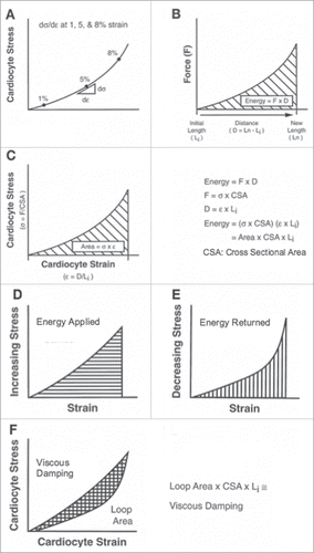
FIGURE 5. Application of the virtual power theory to growth and remodeling of bone and saccular aneurysms. Bone tissue microstructure (A) at different anatomical locations within the femoral head. Used with permission.Citation78-81 Modeling the remodeling process as a rotation of the material axes: (B) states of remodeling equilibrium, adapted with permission,Citation81 (C) evolution of the microstructure to a remodeling equilibrium point for different initial conditions (top) and of the corresponding principal strain ellipsoid (bottom), adapted with permission.Citation80,81
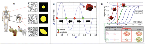
FIGURE 6. Kinematic description of a growing and remodeling cell on a substrate, where the idealized cell can be represented as a collection of material elements (single element depicted as turquoise square). Besides the classical description based on the placement of a body point (x) and of its gradient (Grad x), the evolution of the material microstructure are described by the growth tensor (G) and remodeling variables (m,R). The elastic distorsion tensor is F = (Grad x) G−1.
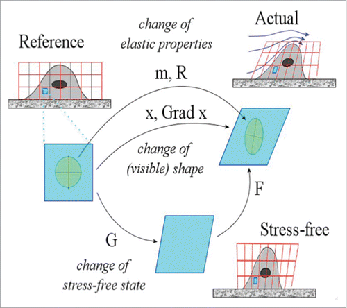
FIGURE 7. Time evolution of the bulk modulus and of its time rate. According to the value of the pair (α,n) parameterizing the external stimulus, this latter can either succeed or fail in triggering cell adaptation to the superimposed pressure.
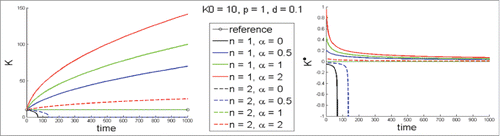
Table 1. Example experimental design to test the theory of stem cell mechanoadaptation. (A) Libraries of mechanical cues will be tested prospectively using independent variables including (1) developmental state, (2) cytoskeletal adaptation, (3) uncoupling of cytoskeletal adaptation from cell shape and volume changes, as well as (4) biochemical controls. (B) Outcome measures will be made on dependent variables including the magnitude, distribution and degree of polymerization of cytoskeletal constituents including, e.g., actin and tubulin, cell and nucleus shape and volume as well as phenotypic measures such as gene expression and ECM protein synthesis. (C) Mechanical properties of the stem cells such as cell and nucleus elastic and shear moduli can be calculated using paired experimental - computation methods such as those demonstrated previously by Song et al.Citation12,20,21