Figures & data
Figure 1. Nuclear degradation occurs during normal homeostasis. Degradation of the nucleus is a part of normal cellular homeostasis in three tissues. Grey nuclei and ND denotes where nuclear degradation occurs in each tissue. A) During the development of the lens, the lens epithelial cells migrate along the lens periphery before flattening out and synthesising crystallins. The middle portion, or nucleus of the lens is devoid of both organelles and the nucleus. B) Keratinocytes proliferate in the basal layer of the epidermis prior to terminal differentiation, where cells come off of the basal lamina and express different structural keratins forming the spinous layer. The nucleus is degraded in the upper layers of the epidermis called the granular layer, prior to the synthesis of the enucleate cornified layer which confers the majority of epidermal barrier function. C) Erythroblasts (red blood cell precursors) are formed by a process of nuclear condensation and extrusion, forming a body called a pyrenocyte, which is engulfed and degraded by adjacent macrophages.
Table 1. Commonalities and differences in the key processes of mammalian nuclear removal; Comparison of known nuclear degradation processes and signalling pathways activated in keratinocytes, lens fibre cells and erythroblasts. A tick denotes that process or phenomenon is active in that cell type, a cross denotes that it is not, and – not determined in that cell type.
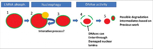
Figure 2. A possible order of events in nuclear degradation in keratinocytes. Possible stages of nuclear degradation based on our and other's data. To begin, the nucleus is intact but is marked by phosphorylation of Lamin A/C (1). This targets an autophagolysosome (LC3-positive/LAMP2-positive body, orange) to that region of the nuclear lamina (2). The autophagolysosome removes some of the nuclear content, reducing nuclear size (3). Steps 1–3 are repeated iteratively until the nuclear lamina is sufficiently damaged to allow ingress of DNases. Then large scale degradation of the nuclear material occurs, potentially concomitant with further degradation of the nuclear lamina (5). Red colour denotes nuclear material, while green denotes the nuclear lamina.