Figures & data
Figure 1. Immunofluorescence images showing PNCs detected by a PTBP1-specific antibody (green) and nucleoli labeled with an anti-UBF antibody (red). Nuclei are visualized by DAPI staining (blue) scale bar, 5 μm.
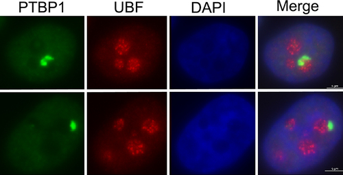
Figure 2. Using the PNC as a readout in metastasis-selective compound screens.
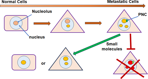
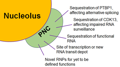
Figure 3. Possible PNC functions in cancer cells.