Figures & data
Figure 1. A photomicrograph of the ovarian structure in the early active stage in female Protothaca (Notochione) jedoensis. Note a number of previtellogenic oocytes (POC) and early vitellogenic oocytes (VOC) containing a few follicle cells (FC, arrow) in the lumina (LU) of the oogenic follicles with follicular walls (FW).
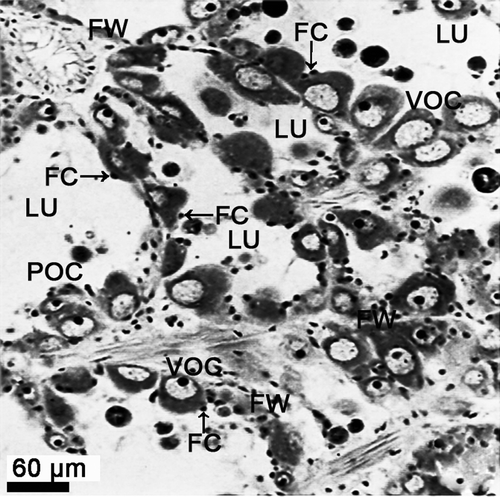
Figure 2. Electron micrograph of oogenesis in female Protothaca (Notochione) jedoensis. Oogonia (OG). Note a large nucleus (N), and several mitochondria (M) and vacuoles in the cytoplasm.
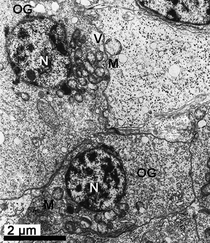
Figure 3. Electron micrograph of oogenesis in female Protothaca (Notochione) jedoensis. A previtellogenic oocyte (PVO) and a follicle cell (FC). Note a nucleolus (NU) in a large nucleus (N), and mitochondria in the cytoplasm of the previtellogenic oocyte (PVO), and well-developed rough endoplasmic reticula (RER, arrow) and glycogen particles (GP) in the cytoplasm of the follicle cell (FC) attached to the oocyte.
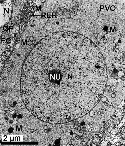
Figure 4. Electron micrograph of oogenesis in female Protothaca (Notochione) jedoensis. An early vitellogenic oocyte (EVO). Note the early vitellogenic oocyte (EVO) containing a large nucleus (N), and several mitochondria (M) and a number of vacuoles (V) in the cytoplasm.
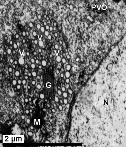
Figure 5. Electron micrograph of oogenesis in female Protothaca (Notochione) jedoensis. An early vitellogenic oocyte (EVO). Note the Golgi product (GPR, arrow) near the vacuoles (V) and vesicles (VE) which is formed by the Golgi complex (G).
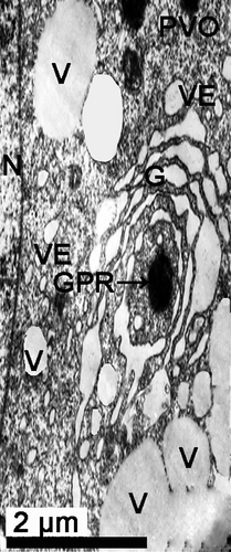
Figure 6. Electron micrograph of oogenesis in female Protothaca (Notochione) jedoensis. An early vitellogenic oocyte (EVO). Note lipid droplets (LD) between well-developed rough endoplasmic reticula (RER) and mitochondria (M) in the cytoplasm of an early vitellogenic oocyte (EVO).
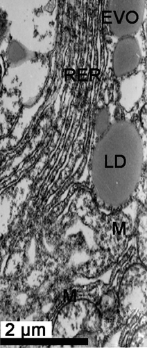
Figure 7. Electron micrograph of oogenesis in female Protothaca (Notochione) jedoensis. An early vitellogenic oocyte (EVO). Note the coated vesicles (CV) occurring through the coated endocytotic pits (CP, arrow) formed by endocytosis at the cortical region near the oolemma (OL).
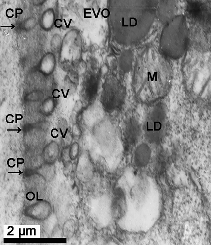
Figure 8. Electron micrograph of oogenesis in female Protothaca (Notochione) jedoensis. A late vitellogenic oocyte (LVO). Note a number of lipid droplets (LD) and glycogen particles (GP) in the cytoplasm near the nucleus (N).
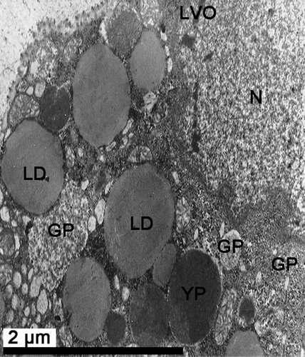
Figure 9. Electron micrograph of oogenesis in female Protothaca (Notochione) jedoensis. A late vitellogenic oocyte (LVO) and follicle cells (FC). Note a number of proteinaceous yolk granules (PYG), lipid droplets (LD), microvilli on the vitellogenic envelope of the oocyte, and follicle cells (FC) containing lipid droplets (LD) and myelin-like organelles (MLO, arrow) in the cytoplasm.
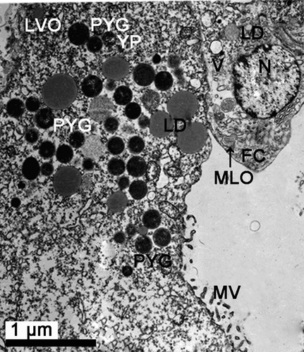
Figure 10. Electron micrograph of oogenesis in female Protothaca (Notochione) jedoensis. A late vitellogenic oocyte (LVO). Note proteinaceous yolk granules (PYG) in the cytoplasm of the oocyte.
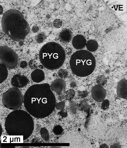
Figure 11. Electron micrograph of a mature oocyte and a degenerating oocyte in female Protothaca (Notochione) jedoensis. (11) Mature yolk granules in a mature oocyte. Note a number of mature yolk granules (MYG) being composed of three parts: crystalline core (CC), electron lucient cortex (ELC), and a limiting membrane (LM, arrow).
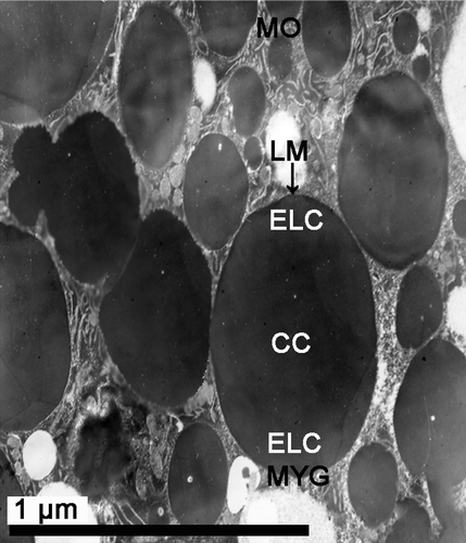
Figure 12. Electron micrograph of a mature oocyte and a degenerating oocyte in female Protothaca (Notochione) jedoensis. The degenerating oocytes (DO). Note a number of degenerating yolk granules (DYG) surrounded with myelin-like organelles (MLO, arrow) in the cytoplasm of the degenerating oocyte (DO).
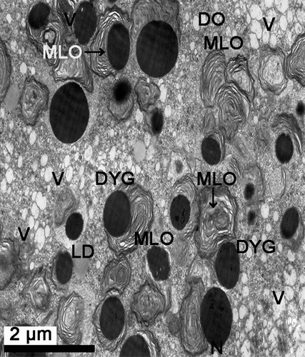
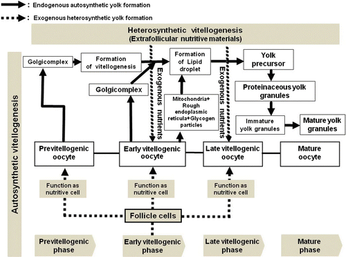
Figure 13. A schematic diagram of the processes of autosynthetic and heterosynthetic vitellogenesis during oogensis in female Protothaca (Notochione) jedoensis.