1,305
Views
4
CrossRef citations to date
0
Altmetric
Articles

Expansion, characterization, and differentiation of rabbit bone marrow-derived mesenchymal stem cells in serum-free medium

, , , , &
Pages 228-236 | Received 20 Jan 2014, Accepted 25 May 2014, Published online: 19 Jun 2014

Figures & data

Table 1. Primers used for RT-PCR and qPCR analyses.

Figure 1. Morphology of primary rabbit bone marrow-derived MSCs. The cells were fibroblastic spindle-shaped at day 4 (A), day 8 (B), and 14 (C) in rbMSCs. Scale bars: 200 µm.
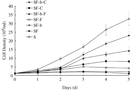
Figure 2. A comparison of the growth of subcultured rbMSCs in the different combination of substrate and growth factor in the defined serum-free medium. Growth curves assay revealed that StemPro® hMSC SFM in the presence of CELLstart and bFGF was suitable for rbMSC proliferation. SF: StemPro® hMSC SFM; C: CELLstart; F: fibronectin; b: bFGF; S: serum.
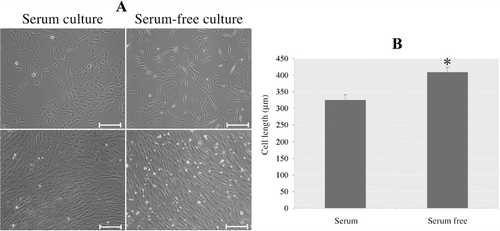
Figure 3. Morphology of subculture of rbMSCs expanded in serum-free and FBS-containing conditions. (A) Phase-contrast image of rbMSCs expanded in FBS and serum-free condition. (B) MSCs cultured in serum-free system presented a longer morphology compared to their counterparts cultured in FBS-containing medium. *P < 0.05. Scale bars: 200 µm.
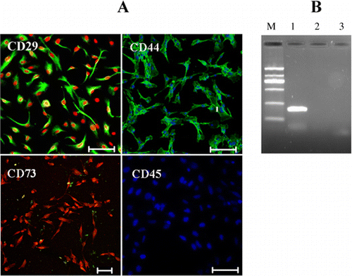
Figure 4. Expression of rbMSC markers in serum-free condition. (A) Immunocytochemical analysis of CD29 (green), CD44 (green), and CD73 (red) expression in rbMSCs cultured in serum-free condition at P1. Hoechst (blue) and PI (red) were stained for cell nuclei. (B) RT-PCR analysis for CD34 (lane 2), CD166 (lane 3), and GAPDH (lane 1). The absence of amplicon in lanes 2 and 3 denoted that these genes were not expressed. M indicates the DNA marker. Scale bars: 200 µm.

Table 2. Quantitative RT-PCR analysis of expanded rbMSCs in both serum-free and FBS containing conditions.
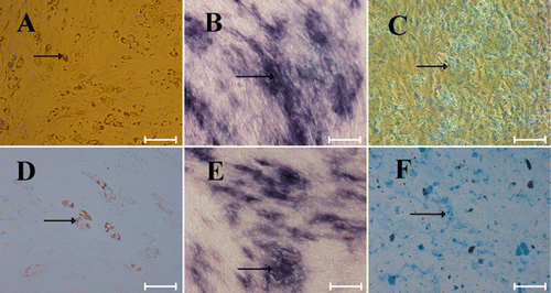
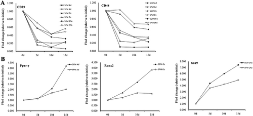
Figure 5. The differentiation ability of rbMSCs grown in FBS-containing (A–C) and serum-free (D–F) media. (A, D) Adipocytic differentiation was detected by Oil-red-O staining (red, arrow); (B, E) osteogenic differentiation was detected by alkaline phosphatase staining (purple, arrow); (C, F) chondrogenic differentiation was detected by alcian blue staining (blue, arrow). Scale bars: 200 µm.
Figure 6. The effect of culture conditions on genes expression in the temporal experiment of trilineage differentiation assay on day 0, day 5, day 10, and day 15. (A) The expression levels of CD29 and CD44 were decreased during differentiation in a time-dependent manner in cells expanded in both FBS and serum-free media. (B) The expression levels of differentiation markers (Ppar-γ, Runx2 and Sox9) were increased in a time-dependent manner in the cells expanded in both FBS and serum-free media. In addition, differentiating cells derived from FBS condition had a lower expression of CD29 and CD44, and a higher expression of Ppar-γ, Runx2 and Sox9. The gene expression was normalized to day 0. X-axis: days (d) after differentiation; Y-axis: fold changes (relative to initial) in gene expression levels.

Reprints and Corporate Permissions

Please note: Selecting permissions does not provide access to the full text of the article, please see our help page How do I view content?

To request a reprint or corporate permissions for this article, please click on the relevant link below:

Academic Permissions

Please note: Selecting permissions does not provide access to the full text of the article, please see our help page How do I view content?

Obtain permissions instantly via Rightslink by clicking on the button below:

If you are unable to obtain permissions via Rightslink, please complete and submit this Permissions form. For more information, please visit our Permissions help page.