615
Views
1
CrossRef citations to date
0
Altmetric
Articles

Production of an epitope-specific antibody using recombinant repetitive oligonucleotides

, , , &
Pages 259-266 | Received 20 Apr 2014, Accepted 02 Jul 2014, Published online: 07 Aug 2014

Figures & data

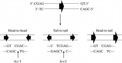
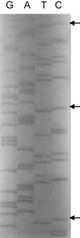
Table 1. Nine amino acids of the human brain β-tubulin type III C-terminus, E. coli codon usage in the target peptide, and the linkers at the 5′- and 3′-ends for ligation of repetitive oligonucleotides.

Figure 1. Construction of pGEX-Acc and pGEX-hβ-tubulin-C9. The pGEX-Acc vector was constructed by insertion of a synthetic oligonucleotide linker into the BamHI and EcoRI sites of pGEX-KT. pGEX-hβ-tubulin-C9 vector for expression of the target peptide was constructed by ligation of the repetitively linked target sequence to a pGEX-Acc vector that had been digested with the AccI restriction enzyme.
Figure 2. The three possible ligation products from the repetitive oligonucleotides. The ligation products in a head-to-tail orientation would be placed in-frame into the vector and thus express the repetitive peptide as a GST-fusion protein when induced in a bacterial system.
Figure 3. AccI digestion of pGEX-KT/pGEX-Acc and synthesis of multiple direct repeats of the target oligonucleotide. A: pGEX-KT was not digested with AccI (Lane 1), but pGEX-Acc was digested by AccI and resulted in a 4.9 kb linear fragment of DNA (Lanes 2 and 3). B: detection of various ligation products, with some larger than 2-kb.
Figure 4. Western blots of total protein isolated from transformed bacterial colonies. Various sizes of repeated peptide were detected using an anti-GST mAb. Lane 1, GST only; Lane 2, 7-unit repeats; Lanes 3, 5, 6, 9; 11-unit repeats; Lane 4, 5-unit repeats; Lane 7, 30-unit repeats; and Lane 8, 10-unit repeats.
Figure 5. DNA sequence determination of the repeated oligonucleotides. The arrow shows a single-unit repeat of the target sequence 5′-CGAGGAAGAATCTGAATCTCAGGGTCCGAAAGT-3′. A pGEX primer and the pGEX-hβ-tubulin-C9 vector were used in the sequencing of the repeated target oligonucleotide.
Figure 6. Purification of fusion proteins and cleavage with thrombin. A: the eluted fusion protein containing the peptide repeats was stained with Coomassie blue dye. Lane M, protein molecule weight marker; Lane 1, GST only; Lane 2, purified fusion protein. B: A 40-kDa fusion protein was detected with the GST antibody. C: the repeated peptide was cleaved by thrombin and released from the 40-kDa fusion protein. Lane 1, 0 min; Lane 2, 30 min; Lane 3, 60 min; and Lane 4, 240 min.
Figure 7. Detection of human brain β-tubulin and the specificity of the generated antibodies. A: total human brain protein was utilised in immunoblot assays with various anti-tubulin Abs including the pAb and mAbs generated throughout this study: 1, commercial mAb to β-tubulin type III; 2, commercial pAb to β-tubulin; 3, pAb to fusion protein (10−1 dilution); 4, mAb (#50) to repeated peptide; 5, mAb (#99) to repeated peptide. Note that pAb and mAbs generated reacted strongly to the 55-kDa human brain β-tubulin. B: total protein extracted from various animal brains (including human) was separated by SDS-PAGE and stained with Coomassie blue dye. C: corresponding immunoblot of gel from Panel B plus positive human β-tubulin control probed with mAb (#50): Lane M, protein molecule weight marker; Lane 1, total protein from human brain; Lane 2, ox; Lane 3, pig; Lane 4, dog; Lane 5, cat; Lane 6, rat; Lane 7, chicken; P, positive human brain β-tubulin III. Note that the mAb specifically reacted to only human brain.

Reprints and Corporate Permissions

Please note: Selecting permissions does not provide access to the full text of the article, please see our help page How do I view content?

To request a reprint or corporate permissions for this article, please click on the relevant link below:

Academic Permissions

Please note: Selecting permissions does not provide access to the full text of the article, please see our help page How do I view content?

Obtain permissions instantly via Rightslink by clicking on the button below:

If you are unable to obtain permissions via Rightslink, please complete and submit this Permissions form. For more information, please visit our Permissions help page.