690
Views
1
CrossRef citations to date
0
Altmetric
Articles

Protease-activated receptor 1-induced GABA release in cultured cortical astrocytes pretreated with GABA is mediated by the Bestrophin-1 channel

&
Pages 244-249 | Received 22 May 2014, Accepted 06 Jul 2014, Published online: 19 Aug 2014

Figures & data

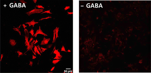
Figure 1. Cortical astrocytes do not contain significant amounts of GABA but can take up extracellular GABA. Representative immunocytochemistry images using anti-GABA antibody in cultured cortical astrocytes with pretreatment by GABA (left) and without GABA pretreatment (right).
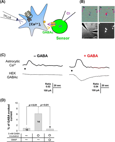
Figure 2. Ca2+-dependent GABA release via the GABA transporter in cultured cortical astrocytes requires pretreatment by GABA. (A) Schematic illustration of sniffer-patch technique. Left pipette, pressure application of TFLLR, a selective agonist of the PAR1 receptor. Right pipette, recording pipette for HEK293T cell expressing GABAC receptors (green). GABA released from cultured astrocyte (blue) upon TFLLR application. On completion of these experiments, GABAC receptors of the sensor cell were fully activated by bath application of GABA (100 µM) so that the response to released GABA could be normalized according to the number of GABA receptors expressed in the sensor cell. Time-dependent reductions in sensor cell currents are due to desensitization of GABAC receptors. (B) Microscope image of sniffer-patch assay. Ca2+ imaging (upper images), GABAC receptor expressed in HEK293T cell and shRNA-transfected astrocyte (lower images). (C) Representative recordings from sniffer-patch assay for each experimental condition (left: normal medium; –GABA; right: pretreatment with GABA; +GABA). Upper trace: Ca2+ transient recorded from astrocyte. Lower trace: whole-cell current recorded from sensor cell (Vh = −70 mV) upon TFLLR pressure application. Diamond: TFLLR application (10 psi, 100 ms, 500 mM). (D) Summary bar graph of GABA release measured under the indicated conditions (SNAP: SNAP5114; GABA transporter inhibitor), with values normalized as described above. Significance was determined by a paired Student's t-test.
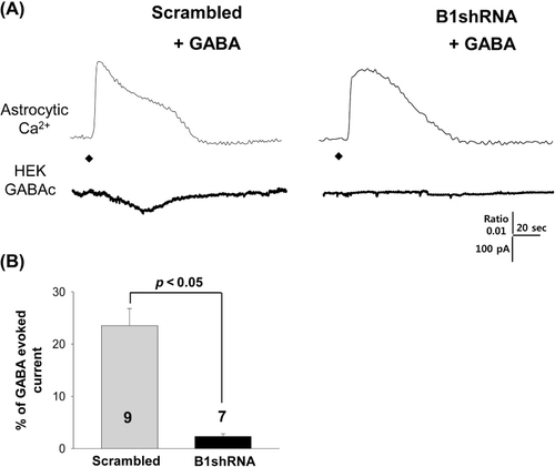
Figure 3. Ca2+-dependent GABA release with GABA pretreatment is mediated by Best1. (A) Representative recordings from sniffer-patch assay for each experimental condition (left: Scrambled shRNA; right: Best1 shRNA). Upper trace: Ca2+ transient recorded from astrocyte. Lower trace: whole-cell current recorded from sensor cell (Vh = −70 mV) upon TFLLR pressure application. Diamond: TFLLR application (10 psi, 100 ms, 500 mM). (B) Summary bar graph of GABA release measured under the conditions indicated, with values normalized as described above. Significance was determined by a paired Student's t-test.

Reprints and Corporate Permissions

Please note: Selecting permissions does not provide access to the full text of the article, please see our help page How do I view content?

To request a reprint or corporate permissions for this article, please click on the relevant link below:

Academic Permissions

Please note: Selecting permissions does not provide access to the full text of the article, please see our help page How do I view content?

Obtain permissions instantly via Rightslink by clicking on the button below:

If you are unable to obtain permissions via Rightslink, please complete and submit this Permissions form. For more information, please visit our Permissions help page.