355
Views
0
CrossRef citations to date
0
Altmetric
Articles

Anthriscus sylvestris-derived extract induces Th1 and Th17 cell differentiation via the upregulation of IL12 and IL23 production

&
Pages 237-243 | Received 20 Jun 2014, Accepted 11 Jul 2014, Published online: 19 Aug 2014

Figures & data

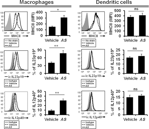
Figure 1. AS-derived extract induces the expression of MHC class II, IL12, and IL23 in macrophages rather than peripheral DCs.Note: DO11.10 Balb/c mice were intraperitoneally injected with OVA peptide323–339 (100 µg) plus either vehicle or AS extract (4 µg) three times a week for two weeks. Splenocytes were prepared from both vehicle- or AS extract-treated mice after immunization. Both the MHC class II cell surface and intracellular IL12p40/IL23p19 production levels were measured on macrophages (gated on F4/80+CD11b+CD11c- population) or DCs (gated on CD11c+ population) by flow cytometry. Data are shown as the means ± SD (n = 3; *P ≤ 0.05; **P ≤ 0.01).
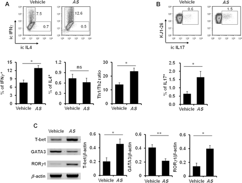
Figure 2. In vivo treatment with AS extracts induces Th1 and Th17 differentiation.Note: After two weeks of immunization, purified splenic CD4+ T cells were activated with plate-bound anti-CD3 (10 µg/ml) and anti-CD28 (1 µg/ml) mAbs for 16 hrs and subsequently restimulated with PMA (50 ng/ml) and ionomycin (1 µg/ml) in the presence of brefeldin A (10 µg/ml) for an additional 2 hrs. (A) The production levels of intracellular IFNγ and IL4 by CD4+ T cells gated on CD4+KJ1-26+ population were measured by flow cytometry. (B) The intracellular IL17 production level by CD4+KJ1-26+ T cells was assessed by flow cytometric analysis. (C) The mRNA levels of T helper cell transcription factors in purified splenic CD4+ T cells from either vehicle- or AS extract-treated mice were analyzed by RT-PCR. Data are shown as the mean ± SD (n = 3; *P ≤ 0.05; **P ≤ 0.01).
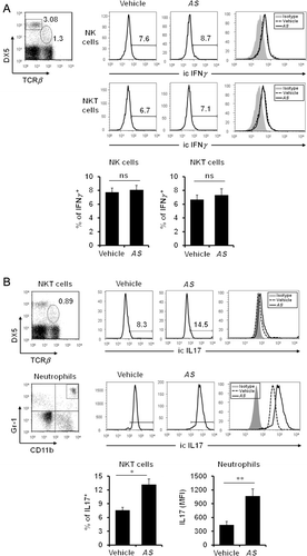
Figure 3. In vivo treatment with AS extracts upregulates IL17 production by NKT cells and neutrophils.Note: After two weeks of immunization, the intracellular expression levels of IFNγ (A) and IL17 (B) by splenocytes from vehicle- or AS extract-treated mice were measured by flow cytometry gated on DX5+TCRβ (NK cells), DX5+TCRβ+ (NKT cells), or non-B/non-T (NBNT) Gr-1+CD11b+ (neutrophils) populations. Data are shown as the means ± SD (n = 3; *P ≤ 0.05; **P ≤ 0.01).
Figure 3. In vivo treatment with AS extracts upregulates IL17 production by NKT cells and neutrophils.Note: After two weeks of immunization, the intracellular expression levels of IFNγ (A) and IL17 (B) by splenocytes from vehicle- or AS extract-treated mice were measured by flow cytometry gated on DX5+TCRβ− (NK cells), DX5+TCRβ+ (NKT cells), or non-B/non-T (NBNT) Gr-1+CD11b+ (neutrophils) populations. Data are shown as the means ± SD (n = 3; *P ≤ 0.05; **P ≤ 0.01).
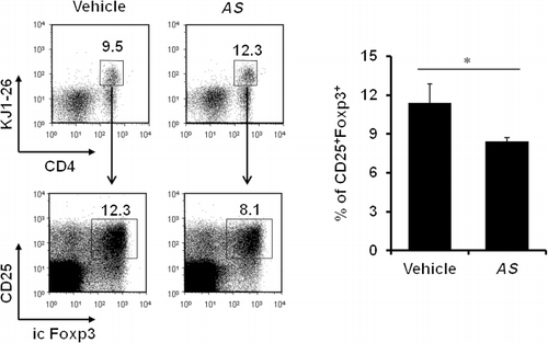
Figure 4. Addition of AS extracts results in a dramatic reduction in the generation of OVA-specific Foxp3+ Treg cells.Note: After two weeks of immunization, total splenocytes from either vehicle- or AS extract-treated mice were prepared and permeabilized for intracellular Foxp3 staining. The intracellular expression of Foxp3 was analyzed by flow cytometry gated on CD4+CD25+KJ1-26+ population. Data are shown as the means ± SD (n = 3; *P ≤ 0.05).

Reprints and Corporate Permissions

Please note: Selecting permissions does not provide access to the full text of the article, please see our help page How do I view content?

To request a reprint or corporate permissions for this article, please click on the relevant link below:

Academic Permissions

Please note: Selecting permissions does not provide access to the full text of the article, please see our help page How do I view content?

Obtain permissions instantly via Rightslink by clicking on the button below:

If you are unable to obtain permissions via Rightslink, please complete and submit this Permissions form. For more information, please visit our Permissions help page.