597
Views
2
CrossRef citations to date
0
Altmetric
Articles

Negative regulation of peroxiredoxin 6 (Prdx 6) transcription by nuclear oncoprotein DEK during leukemia cell differentiation

, , , , , , , & show all
Pages 318-323 | Received 23 Apr 2014, Accepted 29 Jul 2014, Published online: 18 Sep 2014

Figures & data

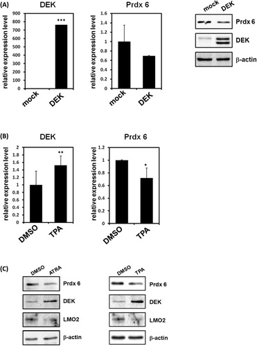
Figure 1. DEK represses Prdx 6 during leukemia cell differentiation. (A) The expression levels of DEK and Prdx 6 in 293T cells with transiently overexpressed DEK were detected via RT-PCR. The error bars represent 2−ΔΔCT ± the SD of three independent experiments. ***P < 0.001. Total proteins were extracts from DEK transfected 293T cells and then were subjected to Western blot analysis using specific antibodies. (B) HL-60 cells were treated with TPA or DMSO. After 48 h, RT-PCR was performed to compare the expression levels of the target genes. Results are shown as means ± SDs; n = 3. **P < 0.01; *P < 0.05. (C) HL-60 cells were treated with ATRA or TPA. After 48 h, differentiated HL-60 cells were lysed and immunoblotted with anti-DEK, anti-β-actin, and anti-Prdx 6 antibodies.
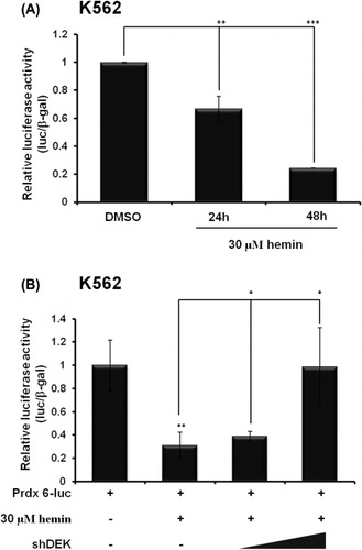
Figure 2. DEK negatively regulates Prdx 6 transcription during leukemia cell differentiation. (A) K562 cells were transfected with Prdx 6-luc and hemin (30 µM) was treated for indicate time points. Cell extracts were assayed for luciferase activity. Luciferase activities were normalized to those of β-galactosidase. Results are shown as means ± SDs; n = 3. **P < 0.01; ***P < 0.001. (B) K562 cells were transfected with Prdx 6-luc and shDEK. Hemin (30 µM) was treated for indicated time points. Cell extracts were assayed for luciferase activity. Luciferase activities were normalized to those of β-galactosidase. Results are shown as means ± SDs; n = 3. **P < 0.01; *P < 0.05.
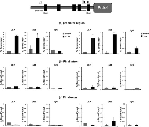
Figure 3. Occupancy of DEK and the p65 subunit in the Prdx 6 promoter region during leukemia cell differentiation. (A) Schematic diagram of the primer pairs used for ChIP analysis. Black boxes represent the Prdx 6 exons. (B) ChIP analyses of the Prdx 6 promoter in ATRA, TPA-treated HL-60 cells were conducted using anti-DEK, and anti-Prdx 6 and examined via RT-PCR. The results are representative of at least three independent experiments (± SD). ***P < 0.001. *P < 0.05.

Reprints and Corporate Permissions

Please note: Selecting permissions does not provide access to the full text of the article, please see our help page How do I view content?

To request a reprint or corporate permissions for this article, please click on the relevant link below:

Academic Permissions

Please note: Selecting permissions does not provide access to the full text of the article, please see our help page How do I view content?

Obtain permissions instantly via Rightslink by clicking on the button below:

If you are unable to obtain permissions via Rightslink, please complete and submit this Permissions form. For more information, please visit our Permissions help page.