544
Views
0
CrossRef citations to date
0
Altmetric
Articles

Role of the intracellular juxtamembrane domain of discoidin domain receptor 2 in focal adhesion formation

, , , , , & show all
Pages 372-379 | Received 01 Sep 2014, Accepted 22 Sep 2014, Published online: 10 Nov 2014

Figures & data

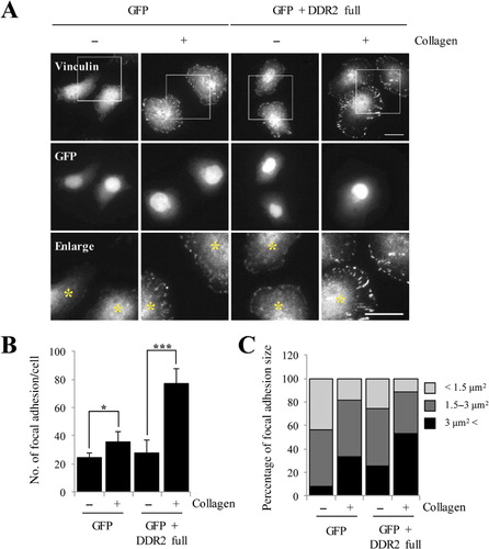
Figure 1. DDR2 activation induced by collagen regulates focal adhesion formation in HeLa cells. (A) HeLa cells were transfected with either GFP alone or GFP and DDR2 together for 24 h. Cells were serum starved for 16 h and then treated with 50 µg/mL Type I collagen for 2 h. Following incubation, samples were fixed and stained using anti-vinculin to visualize focal adhesion formation. Scale bar: 20 µm. (B) Data represent the mean and SD (error bars) in n = 20 cells for each condition. (C) Focal adhesion size was measured and grouped into three categories (<1.5 µm2, 1.5–3 µm2, and >3 µm2). A significant increase in both size and number of focal adhesion was observed when DDR2 is expressed. *P < 0.05, ***P < 0.001, Student's t-test.
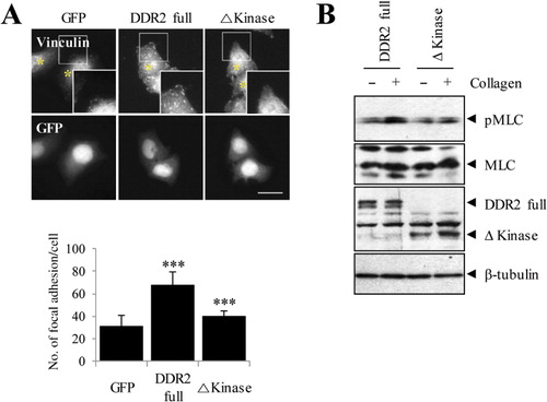
Figure 2. Inhibition of DDR2 activation reduces formation of focal adhesion. (A) HeLa cells were expressed with designated plasmid for 24 h. After 16 h serum starvation, cells were treated with 50 µg/mL Type I collagen for 2 h and stained using an anti-vinculin antibody to visualize focal adhesions. (B) HeLa cells transfected with full and Δkinase DDR2 constructs were lysed and subjected to SDS-PAGE followed by anti-phospho-MLC (serine 19), anti-pan-MLC, anti-myc, and anti-β-tubulin antibodies. Cells that expressed Δkinase DDR2 inhibited the phosphorylation of MLC induced by collagen stimulation. ***P < 0.001, Student's t-test.
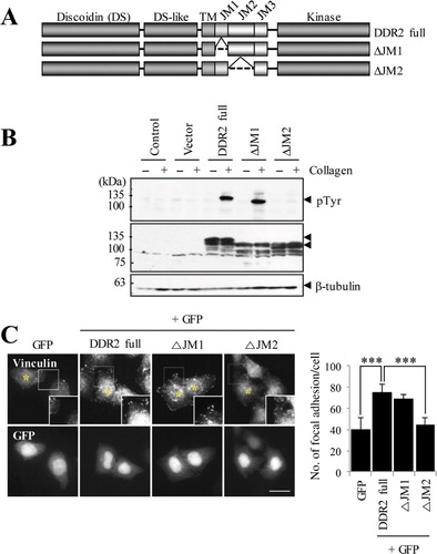
Figure 3. DDR2 activation via the JM2 domain is involved in focal adhesion formation. (A) Schematic diagram of DDR2 truncated mutants. (B) HEK293 cells were transfected with plasmids for 36 h. Cells were lysed, separated on SDS-PAGE and then subjected to western blotting with anti-phophotyrosine (PY20), anti-myc, and anti-β-tubulin antibodies. (C) HeLa cells were transfected with DDR2 mutants and GFP simultaneously (5:1 molar ratio) for 24 h. Following incubation, cells were fixed and stained with an anti-vinculin antibody to visualize focal adhesions. Scale bar: 20 µm. Data (right-hand side) indicate the number of focal adhesion in each condition (n = 10). ***P < 0.001.
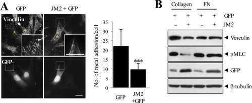
Figure 4. JM2 region exerts dominant-negative effects on focal adhesion formation in MEF cells. (A) MEF cells were transiently transfected with either GFP only or GFP and myc-tagged isolated JM2 (1:5 molar ratio) for 24 h, after which cells were serum starved for 16 h and treated with 50 µg/mL Type I collagen for 2 h. Following incubation, cells were fixed and stained with anti-vinculin antibody. Asterisks indicate transfected cells. Scale bar: 20 µm. (B) Transfected MEFs were seeded on a 35-mm dish and serum starved for 16 h. Treatments of Type I collagen (50 µg/mL) and human plasma fibronectin (10 µg/mL) were then applied for 2 h. Following collagen stimulation, MEF cells expressing JM2 showed reduced MLC phosphorylation. ***P < 0.001, Student's t-test.
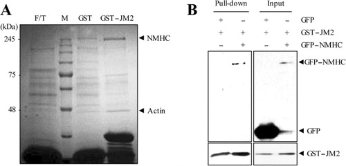
Figure 5. JM2 domain interacts with nonmuscle myosin II (NMHC). (A) After chromatography of mouse liver lysates on sepharose beads bound to the GST or GST-JM2 protein, eluted proteins were visualized by silver staining. The major binding proteins were analyzed by MALDI-TOF mass spectrometry. The results of mass spectrometry are indicated to the right of the panel. (B) HEK 293T cells were transfected with either GFP or GFP-tagged nonmuscle myosin II. Total cell lysates were incubated with GST-JM2 protein for 4 h. The pull-down proteins with glutathione sepharose 4B were resolved on SDS-PAGE and analyzed with anti-GFP antibody. These results showed that JM2 domain of DDR2 specifically and directly interacts with myosin.

Reprints and Corporate Permissions

Please note: Selecting permissions does not provide access to the full text of the article, please see our help page How do I view content?

To request a reprint or corporate permissions for this article, please click on the relevant link below:

Academic Permissions

Please note: Selecting permissions does not provide access to the full text of the article, please see our help page How do I view content?

Obtain permissions instantly via Rightslink by clicking on the button below:

If you are unable to obtain permissions via Rightslink, please complete and submit this Permissions form. For more information, please visit our Permissions help page.