Figures & data
Figure 1. Resveratrol inhibits prostate cancer cells. (a) The molecular structure of resveratrol. (b) Cell proliferation was analyzed with the MTT assay. (c) Quantitative results of PC-3 cell migration. (d) Cell migration was evaluated using a wound-healing assay. Representative images are shown at 0 and 24 h post-wounding with ×40 magnification. Scale bars = 500 μm. Statistical significance was assessed by one-way ANOVA.
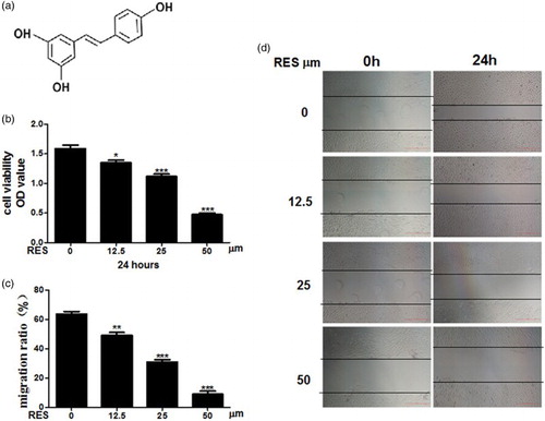
Figure 2. Resveratrol induces apoptosis in prostate cancer cells. (a) Apoptosis was determined by Annexin V/PI double staining followed by flow cytometry. (b) Nucleic morphology were stained with Hoechst 33258; the white arrows represent location of apoptosis cells. (c) Flow cytometry analysis of the cell cycle in PC-3 cells and quantification of the number of cells in G0/G1, S and G2/M phases. (d, e) Real-time PCR and Western blot analyses of the apoptosis-related proteins. (f) Results of the ΔΨm test in the cells, ×200 magnification. Scale bars = 100 μm. Statistical significance was assessed by the unpaired Student's t-test.
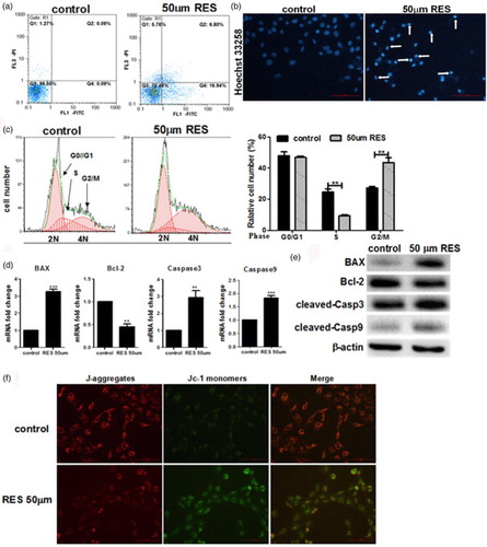
Figure 3. Resveratrol inhibits the EMT in prostate cancer cells. (a) Real-time PCR and Western blot analyses of the expression of E-cadherin and Vimentin in the cells. (b) Immunofluorescence detection of the E-cadherin and vimentin proteins in the PC-3 cells at ×200 magnification. Green and red indicates the protein of interest. Scale bars = 100 μm. (c) Quantification of E-cadherin and Vimentin expression using Image-Pro Plus 6.0 software. Statistical significance was assessed by the unpaired Student's t-test.
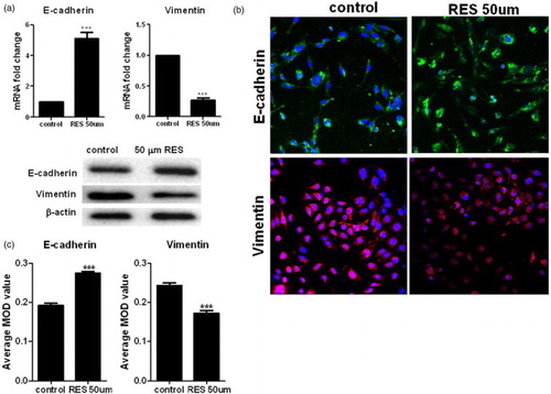
Figure 4. Roles of resveratrol in the phosphorylation of Akt in prostate cancer cells. Western blot analysis of the expression of p-Akt and Akt in the cells. Statistical significance was assessed by one-way ANOVA.

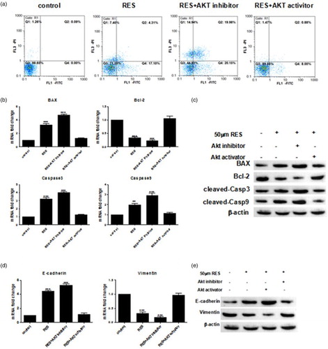
Figure 5. Resveratrol promotes apoptosis and suppresses the EMT in PC-3 cells by suppressing the PI3K/Akt signaling pathway. (a) Apoptosis was determined by Annexin V/PI double staining followed by flow cytometry. (b, d) Real-time PCR and western blot analyses of the apoptosis-related proteins. (c, e) Real-time PCR and Western blot analyses of the EMT-related proteins. Statistical significance was assessed by one-way ANOVA.