Figures & data
Figure 1. Different stages of spermatogenesis observed in the testis of L. cornutus. Spermatogenesis of L. cornutus consists of four distinctive stages: spermatogonium, spermatocyte, spermatid, and spermatozoon. SG: silk gland; Ts: Testis. White arrowhead indicates spermatogonia; Black arrowhead indicates spermatocytes; Black arrow indicates spermatids undergoing spermiogenesis. White arrow indicates spermatozoon. Magnifications are as follow: (a) ×40; (b) ×100; (c) ×200; (d) ×400.
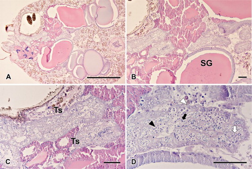
Figure 2. Transverse section of the testis in L. cornutus. Spermatogenesis in spider testis occurs along inward direction. Spermatogonium stays along the testicular membrane, maintaining the cell line. As development progresses, spermatocytes moves inward to produce differentiated cells. SG: spermatogonium; SCp: primary spermatocyte; SCs: secondary spermatocyte; ST: spermatid. Magnifications are as follow: (a, b, e) ×2800; (c, d) ×3900; (f): ×5800.
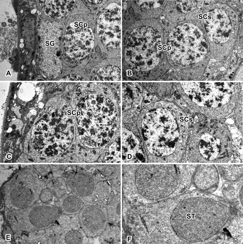
Table 1. Numerical data about germ cell morphology.
Figure 3. Image editing steps prior to rendering. (a) Light microscopic sectioned images with basic H&E staining. (b) Images converted into grayscale mode due to sensitivity of the program. (c) Unwanted portions may be trimmed to increase accuracy. (d) Color inversion completed in order to eliminate background in the rendering program.
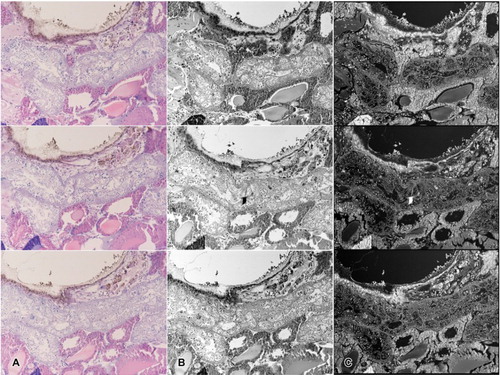
Figure 4. Image loading steps using the Amira program. (a) Set of images loaded in Amira. When the axes are aligned, serial images can be viewed through the Orthoslices tool. (b) Image labeled using the Segmentation editor. Yellow colored area indicates a labeled cyst. Spermatocytes were labeled separately so as not to interrupt the channels. (c–d) Images before applying the Alignslices tool (c). Arrowhead indicates unaligned portion of the image. Aligned images after applying the Alignslices tool (d).
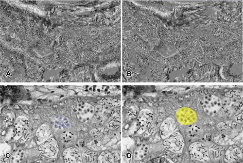
Figure 5. Testicular cyst rendered into 3D image. (a) Images were taken while rotating every 30 degrees on the XY axis. (b) Images were taken while rotating every 30 degrees on the YZ axis. (c) Images were taken while rotating every 30 degrees on the XZ axis.
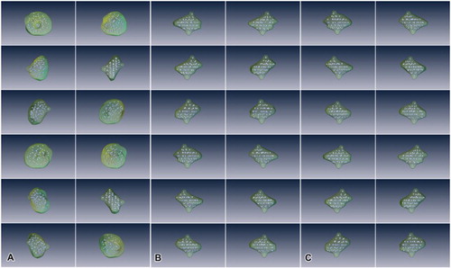
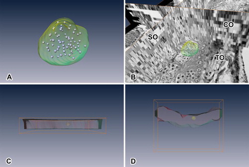
Figure 6. Rendered image viewed in different mode and angle. (a) Rendered cyst and spermatocytes in comparison with the images used. TO indicates transverse orthoslice, SO indicates sagittal orthoslice, CO indicates coronal orthoslice. SO and CO are produced after image alignment. (b) Lateral view of rendered testicular cyst with testis along the XZ axis. (c) Lateral view of rendered testicular cyst with testis along the XY axis. (d) Lateral view of rendered testicular cyst with testis along the YZ axis. *Rendered red portion indicates comparative volume of testis used in the reconstruction.