
Figures & data
Figure 1. Heat stress induced ferroptosis-like death in GMECs. One-year-old normal healthy adult goats in experimental group or control group were raised in an environment of 40°C ± 2°C or 20°C ± 2°C for 4 weeks and then slaughtered, (A) each tissue was taken and the selenium content was determined using the ID-HG-ICP-MS method. The DMECs isolated from 1-year-old Saanen dairy goats at the peak lactation (15 days postpartum) were used for heat stress treatment, then the cell viability, iron ion concentration and ROS level were measured to determine the extent of ferroptosis: (B) MTT assay was used to detect the cell viability. (C) Determination of ROS content. (D) Determination of iron ion concentration by spectrophotometer. (E) RT-PCR was used to detect the expression of GPX4 nucleic acid. (F) Western blotting was used to detect the expression of GPX4 protein. (G) SOD determination kit was used to determine the activity of SOD. *P < 0.05 versus control, **P < 0.01 versus control, ***P < 0.001 versus control, n=6.
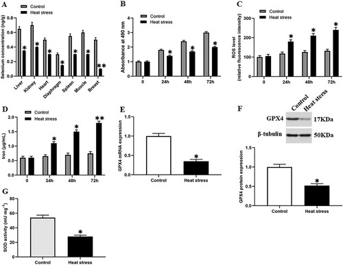
Figure 2. Selenium incubation reduces heat stress-induced ferroptosis-like death of GMECs. After adding a certain concentration of selenite (1, 5, 10, 20 μM) to heat-stressed GMECs for 24 h, the cell viability, iron ion concentration and ROS level were measured: (A) The negative control group was incubated with different concentrations of selenium to test the effect of selenium addition on cell viability. (B) MTT assay was used to detect the cell viability. (C) Determination of ROS content. (D) Determination of iron ion concentration by spectrophotometer. *P < 0.05 versus control, **P < 0.01 versus control, n=6.
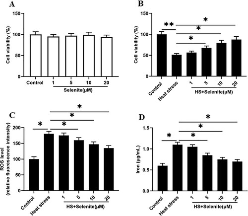
Figure 3. Selenium incubation improves GPX4 expression and SOD activity under heat stress. After adding a certain concentration of selenite (1, 5, 10, 20 μM) to heat-stressed GMECs for 24 h, the expression of GPX4 and the activity of SOD were detected: (A) RT-PCR was used to detect the expression of GPX4 nucleic acid. (B) Western blotting was used to measure the expression of GPX4 protein. (C) SOD determination kit was used to determine the activity of SOD. *P < 0.05 versus control, **P < 0.01 versus control, n=6.
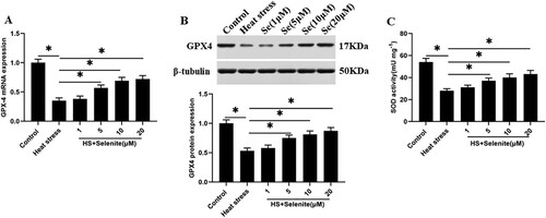
Figure 4. SOD activation reduces heat stress-induced ferroptosis-like death of GMECs. Adding a certain concentration of SOD activator CuCl2 (15, 30 mM) to heat-stressed GMECs. After 24 h of co-cultivation, the degree of cell ferroptosis-like death can be judged by detecting cell viability, iron ion concentration, and ROS level: (A) SOD determination kit was used to determine the activity of SOD. (B) Western blotting was used to measure the expression of GPX4 protein. (C) MTT assay was used to detect the cell viability. (D) Determination of ROS content. (E) Determination of iron ion concentration by spectrophotometer. *P < 0.05 versus control, n=6.
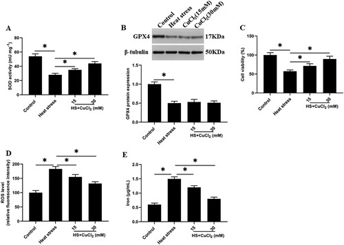
Figure 5. Knockdown of GPX4 inhibits SOD activity and increases ferroptosis-like death of GMECs. GMECs cultured in DMEM were treated by different concentrations of GPX4 siRNA (20, 40 nM), then cells and supernatants were isolated for functional determinations: (A) RT-PCR was used to determine the expression of GPX4 nucleic acid. (B) Western blotting was used to measure the expression of GPX4 protein. (C) GMECs were treated with 40 nM GPX4 siRNA and 15 mM CuCl2, cultured for 24 h, and then SOD determination kit was used to determine the activity of SOD. (D) MTT assay was used to detect the cell viability. (E) Determination of ROS content. (F) A spectrophotometer was used to determine the concentration of iron ions. *P < 0.05 versus control, n=6.
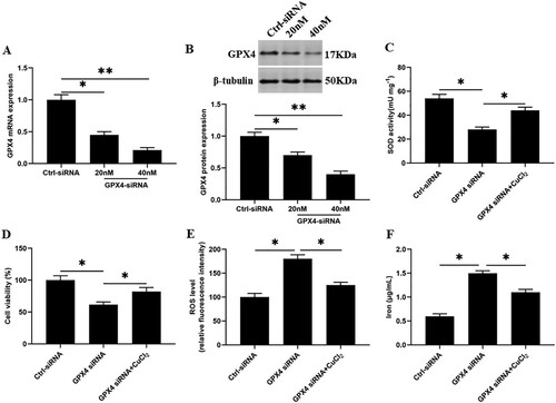
Figure 6. Overexpression of GPX4 reduces SOD activity and reduces heat stress-induced ferroptosis-like death in GMECs. The negative control group and heat stimulation group were transfected with 1 mg/mL adenovirus carrying GPX4, and then the cells and supernatant were separated for functional testing: (A) RT-PCR was used to determine the expression of GPX4 nucleic acid. (B) Western blotting was used to measure the expression of GPX4 protein. (C) SOD determination kit was used to determine the activity of SOD. (D) MTT assay was used to detect the cell viability. (E) Determination of ROS content. (F) A spectrophotometer was used to determine the concentration of iron ions. *P < 0.05 versus control, n=6.
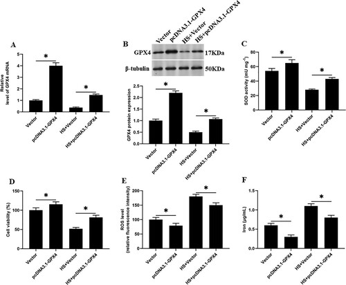
Figure 7. The activation of mTOR inhibits the expression of GPX4 and the activity of SOD. Adding 10 μM selenite or mTOR activator MHY1485 to heat-stressed GMECs. After 24 h of culture, the cells and supernatant were separated for functional determination: (A) mTOR determination kit was used to determine the activity of mTOR. (B) Western blotting was used to measure the expression of GPX4 protein. (C) SOD determination kit was used to determine the activity of SOD. (D) MTT assay was used to detect the cell viability. (E) Determination of ROS content. (F) A spectrophotometer was used to determine the concentration of iron ions. *P < 0.05 versus control, **P < 0.01 versus control, n=6.
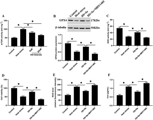
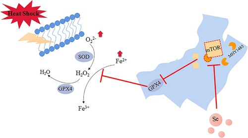
Figure 8. Selenium promotes the expression of GPX4 by inhibiting the activity of mTOR. Under heat stress treatment, the iron ion concentration and ROS level of GMECs increased significantly and caused ferroptosis-like death in the cells. The activity of mTOR was higher under heat stress, which further inhibited the expression of GPX4, the addition of selenium inhibited the activity of mTOR under heat stress and improved the expression of GPX4, and then the activity of SOD is increased, thereby improving ferroptosis-like death under heat stress.
Supplemental Material
Download TIFF Image (12.3 MB)Data availability statement
All data generated or analyzed during this study are included in this published article.