
Figures & data
Table 1. Distribution of Intracranial Tumours in Enugu [2008–2017].
Table 2. Age and sex distribution of pituitary adenomas in Enugu [2008–2017].
Figure 1. Histologic patterns of pituitary adenomas in Enugu (2008–2017)..
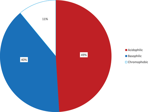
Figure 2. Distribution of immunohistochemical subtypes of pituitary adenomas by sex in Enugu (2008–2017).
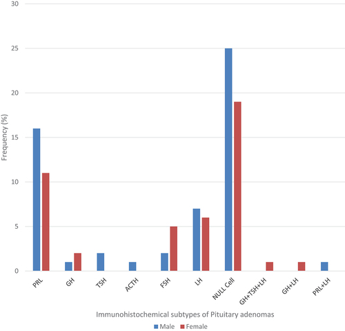
Figure 3. Photomicrographs of pituitary adenomas showing (a) acidophilic, (b) basophilic and (c) chromophobic adenomas [H&E stain x 400].
![Figure 3. Photomicrographs of pituitary adenomas showing (a) acidophilic, (b) basophilic and (c) chromophobic adenomas [H&E stain x 400].](/cms/asset/b46b97d2-8581-4ad6-a98a-49cad1ec3f0e/zljm_a_2245587_f0003_oc.jpg)
Figure 4. Photomicrographs of pituitary tissue showing (a) positive cytoplasmic staining with growth hormone monoclonal antibody in normal pituitary gland [Positive GH Immunostain x 400], (b) positive cytoplasmic staining with growth hormone monoclonal antibody in pituitary adenoma [Positive GH Immunostain x 400], (c) positive cytoplasmic staining with prolactin hormone monoclonal antibody in pituitary adenoma [Prolactin Immunostain x 400] and (d) negative cytoplasmic staining in pituitary adenoma [Negative immunostain for all six antibodies x 400].
![Figure 4. Photomicrographs of pituitary tissue showing (a) positive cytoplasmic staining with growth hormone monoclonal antibody in normal pituitary gland [Positive GH Immunostain x 400], (b) positive cytoplasmic staining with growth hormone monoclonal antibody in pituitary adenoma [Positive GH Immunostain x 400], (c) positive cytoplasmic staining with prolactin hormone monoclonal antibody in pituitary adenoma [Prolactin Immunostain x 400] and (d) negative cytoplasmic staining in pituitary adenoma [Negative immunostain for all six antibodies x 400].](/cms/asset/0567fcfd-f561-4aa2-a732-68481f4b10d5/zljm_a_2245587_f0004_oc.jpg)
Figure 5. Photomicrographs of pituitary tissue showing (a) positive cytoplasmic staining with Thyroid Stimulating Hormone Monoclonal Antibody [TSH Immunostain x 400] (b) positive cytoplasmic staining with Adrenocorticotrophic Hormone Monoclonal Antibody [ACTH Immunostain x 400], (c) positive cytoplasmic staining with Follicle Stimulating Hormone Monoclonal Antibody [FSH Immunostain x 400] and (d) positive cytoplasmic staining with Luteinizing Hormone Monoclonal Antibody [LH Immunostain x 400].
![Figure 5. Photomicrographs of pituitary tissue showing (a) positive cytoplasmic staining with Thyroid Stimulating Hormone Monoclonal Antibody [TSH Immunostain x 400] (b) positive cytoplasmic staining with Adrenocorticotrophic Hormone Monoclonal Antibody [ACTH Immunostain x 400], (c) positive cytoplasmic staining with Follicle Stimulating Hormone Monoclonal Antibody [FSH Immunostain x 400] and (d) positive cytoplasmic staining with Luteinizing Hormone Monoclonal Antibody [LH Immunostain x 400].](/cms/asset/324f1f00-d9c9-4ed5-8702-f97de58d26c7/zljm_a_2245587_f0005_oc.jpg)
Table 3. Immunohistochemical subtypes of Pituitary Adenomas in Enugu, 2008–2017.
Table 4. Distribution of Immunohistochemical Subtypes of Pituitary Adenomas by Age, Sex and Biopsy Size in Enugu [2008–2017].