
Figures & data
Figure 1. Analysis of antigen preparations from A. actinomycetemcomitans (A.a.) and P. gingivalis (P.g.) as used for coating of ELISA plates.
Lanes of sodium dodecyl sulfate polyacrylamide (7%) gels were loaded with amounts of antigens equal to those used for coating of one 96-well plate. Staining with Coomassie Blue (polysaccharides and LPS) are not visible. Mr markers in kilo Daltons.
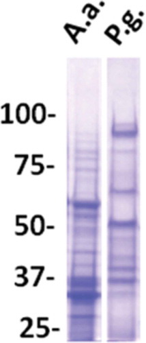
Figure 2. Validation of assay sensitivity and specificity to antigen preparations from A. actinomycetemcomitans.
A) Homologous titration curves for serum from a patient with aggressive periodontitis colonized with the JP2 clone (serotype b) of A. actinomycetemcomitans in wells saturated with disintegrated serotype b bacterial antigen (●) and whole, formalin-treated serotype b bacteria (■), respectively. B) Comparative titration of sera from four microbiologically and clinically characterized individuals in an ELISA plate coated with an equivalent mixture of antigens from three strains of A. actinomycetemcomitans representing serotypes a, b, and c. The four sera were from a patient with aggressive periodontitis colonized with the JP2 clone of A. actinomycetemcomitans (●), a healthy individual colonized by a non-JP2 clone of A. a. (∎), two healthy individuals without detectable A.a. (▲,▼). C) Comparative titrations of a serum from a patient with aggressive periodontitis colonized with the JP2 clone (serotype b) of A. actinomycetemcomitans in ELISA wells coated with antigens from either A. actinomycetemcomitans serotype b (●), serotype a (∎), or serotype c (▲).
Table 1. Distribution of study population exposure- and covariables by cardiovascular disease (CVD) and periodontitis.
Table 2. Periodontitis: univariable logistic regression analyses.
Table 3. Periodontitis: multivariable logistic regression analyses.
Table 4. Cardiovascular disease: univariable logistic regression analyses.
Table 5. Cardiovascular disease: multivariable logistic regression analyses.