2,729
Views
6
CrossRef citations to date
0
Altmetric
Review Article

Innovative application of nested PCR for detection of Porphyromonas gingivalis in human highly calcified atherothrombotic plaques

, , , , , & show all
Article: 1742523 | Received 28 Jul 2020, Accepted 14 Oct 2020, Published online: 08 Apr 2020

Figures & data

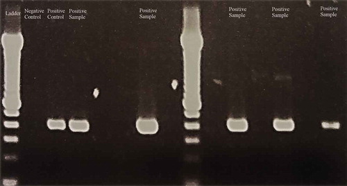
Table 1. Identification of periodontal pathogens in atherothrombotic samples using PCR methods

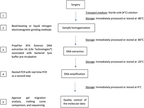
Table 2. Vascular sample preparation for identifying periodontal pathogens

Table 3. PCR conditions and identification methods for periodontal pathogens in atherothrombotic samples

Figure 1. Proposed protocol for Porphyromonas gingivalis 16S rDNA amplification in human calcified atherothrombotic samples in 5 steps. Sterile equipment must be used for all the following steps. (1.) Samples must be transported to the laboratory in a cold (4°C) sterile solution within a few hours after harvesting. (2.) Then, samples must be processed with homogenization. A liquid nitrogen electromagnetic grinding method is preferred over a bead-beating method to maximize nucleic acid preservation. Samples can be stored at −80°C while waiting for homogenization, but freezing and thawing steps should be minimized. (3.) DNA extraction is carried out after pre-incubation with a bacterial lysis buffer using the PrepFiler BTA forensic DNA extraction kit (Life Technologies®) to remove calcifications. (4.) Then, nested PCR is carried out, with conventional PCR as the first step and real-time PCR as the second step (≤40 cycles). (5.) For quality control, the molecular data is obtained using agarose gel migration, melting curve comparison, and sequencing

Figure 1. Proposed protocol for Porphyromonas gingivalis 16S rDNA amplification in human calcified atherothrombotic samples in 5 steps. Sterile equipment must be used for all the following steps. (1.) Samples must be transported to the laboratory in a cold (4°C) sterile solution within a few hours after harvesting. (2.) Then, samples must be processed with homogenization. A liquid nitrogen electromagnetic grinding method is preferred over a bead-beating method to maximize nucleic acid preservation. Samples can be stored at −80°C while waiting for homogenization, but freezing and thawing steps should be minimized. (3.) DNA extraction is carried out after pre-incubation with a bacterial lysis buffer using the PrepFiler BTA forensic DNA extraction kit (Life Technologies®) to remove calcifications. (4.) Then, nested PCR is carried out, with conventional PCR as the first step and real-time PCR as the second step (≤40 cycles). (5.) For quality control, the molecular data is obtained using agarose gel migration, melting curve comparison, and sequencing

Figure 2. Images of agarose gels showing the migration of amplicons from an amplification by direct real-time PCR using primers designed for Porphromonas gingivalis. (a) PCR protocol using 40 cycles. (Lane 1:) Negative control. (Lane 2:) Positive control. (Lanes 3–13 and 15, 16:) Samples. (Lane 14:) Ladder. Only the last sample (lane 16) had an amplicon with the same molecular weight as the positive control. (b) PCR protocol using 50 cycles. (Lane 1:) Positive control. (Lanes 2–10 and 12:) Samples. (Lane 11:) Ladder. (Lane 13:) Negative control. All the samples present multiple amplicons

Figure 2. Images of agarose gels showing the migration of amplicons from an amplification by direct real-time PCR using primers designed for Porphromonas gingivalis. (a) PCR protocol using 40 cycles. (Lane 1:) Negative control. (Lane 2:) Positive control. (Lanes 3–13 and 15, 16:) Samples. (Lane 14:) Ladder. Only the last sample (lane 16) had an amplicon with the same molecular weight as the positive control. (b) PCR protocol using 50 cycles. (Lane 1:) Positive control. (Lanes 2–10 and 12:) Samples. (Lane 11:) Ladder. (Lane 13:) Negative control. All the samples present multiple amplicons

Figure 3. Images of agarose gel showing the migration of amplicons from an amplification by nested PCR (conventional PCR as the first step and real-time PCR as the second step) using primers designed for Porphromonas gingivalis. (Lane 1 and 9:) Ladder. (Lane 2:) Negative control. (Lane 3:) Positive control. (Lanes 4–8 and 10–15:) Samples. Samples from (lanes 4, 7, 11, 13, and 15) had an amplicon with the same molecular weight as the positive control. They were confirmed to be positive by comparison of the melting curves and by sequencing

Figure 3. Images of agarose gel showing the migration of amplicons from an amplification by nested PCR (conventional PCR as the first step and real-time PCR as the second step) using primers designed for Porphromonas gingivalis. (Lane 1 and 9:) Ladder. (Lane 2:) Negative control. (Lane 3:) Positive control. (Lanes 4–8 and 10–15:) Samples. Samples from (lanes 4, 7, 11, 13, and 15) had an amplicon with the same molecular weight as the positive control. They were confirmed to be positive by comparison of the melting curves and by sequencing
Supplemental material

Supplemental Material

Download Zip (5.5 MB)