Figures & data
Figure 1. Enhanced serum survival of A. actinomycetemcomitans strain D7SS ompA1 ompA2 upon supplementation of OMVs. A. actinomycetemcomitans cells were incubated in 50% normal human serum (NHS) at 37°C for 1 h. The assay was performed in the absence or presence of 20, 100, and 200 μg of OMVs, respectively, as indicated. Bacterial serum survival was determined by viable count, and shown is the increase (fold change) in survival relative to incubations in vesicle-free controls (50% NHS in PBS). Supplemented OMVs were obtained from the wild-type strain D7SS (blue squares), and D7SS ompA1 ompA2 (red squares), respectively. Shown are means ± SEM from four independent experiments. *P < 0.05 vs control. **P < 0.05 vs supplementation of 20 μg of OMVs. P > 0.05, D7SS OMVs vs D7SS ompA1 ompA2 OMVs
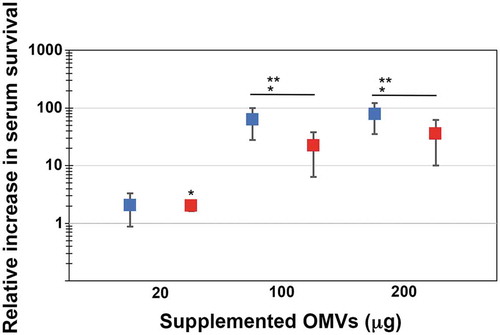
Figure 2. LPS-dependent serum protection of A. actinomycetemcomitans strain D7SS ompA1 ompA2 upon supplementation of OMVs. A. actinomycetemcomitans cells were incubated in 50% normal human serum (NHS) at 37°C for 1 h. The assay was performed in the absence or presence of 20, 100, and 200 μg of OMVs, respectively, as indicated. Bacterial serum survival was determined by viable count, and shown is the increase (fold change) in survival relative to incubations in vesicle-free controls (50% NHS in PBS). Supplemented OMVs were obtained from strain SA3138 (serotype a, wildtype; blue squares), and SA3139 (as SA3138 but LPS lacks the O-antigen polysaccharide part; red squares). Shown are means ± SEM from four independent experiments. *P < 0.05 SA3138 OMVs vs SA3139 OMVs
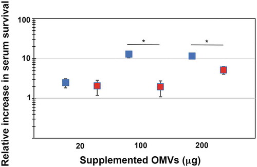
Table 1. Remaining complement activity (%) in the indicated activation pathways after consumption of complement in NHS by A. actinomycetemcomitans OMVs (20 μg; rendering a final OMV protein concentration of 95.2 μg/ml) for 1 h at 37°C. NHS incubated with PBS served as a negative control. Complement consumption was thereafter determined using the COMPL300 kit, as described in Materials and methods. Values are given as mean percentage (range) from two independent experiments
Table 2. Remaining complement activity (%) in the indicated activation pathways after consumption of complement in NHS by A. actinomycetemcomitans LPS (50 ng; 500 EU) for 1 h at 37°C. Complement consumption was thereafter determined using the COMPL300 kit, as described in Materials and methods. Values are given as mean percentage (range) from two independent experiments. P < 0.05 SA3138 OMVs vs SA3139 OMVs for all three tested pathways
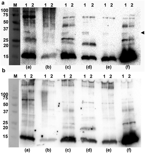
Figure 3. Immunoreactivity of normal human sera to A. actinomycetemcomitans and E. coli OMVs. Western blot analysis of reactivity of NHS from periodontally healthy individuals with (a) OMVs obtained from the A. actinomycetemcomitans strains D7SS (wild-type; lane 1), and D7SS ompA1 ompA2 (lane 2), and (b) OMVs obtained from the A. actinomycetemcomitans strains SA3138 (wild-type; lane 1), and SA3139 (LPS lacks the O-antigen polysaccharide part; lane 2). The NHS samples a-f were used for immunodetection as indicated, and include sera known to exhibit high (a, and f) and low (b) reactivity, respectively, towards recombinant A. actinomycetemcomitans leukotoxin, and one from a confirmed A. actinomycetemcomitans-negative individual (e). Samples equal to 10 μg protein were applied on the gels. The reactive band corresponding to A. actinomycetemcomitans OmpA1 is indicated with an arrowhead. LPS is detected as a diffuse, high-molecular smear pattern for serum sample (b), when assessing SA3138 OMVs. The sizes (kDa) of the proteins in the pre-stained molecular weight marker (M) are indicated along the left sides