Figures & data
Table 1. C. albicans strains used in this study
Table 2. Plasmids used in this study
Table 3. Primers used for BGL2, ECM33, and ALS1 deletion and reintegration
Table 4. Primers used for qRT-PCR
Figure 1. Correlation between NR fluorescence intensity and C. albicans cell concentration. Cell concentrations were determined by measuring the optical density at 600 nm using an ASV11D spectrophotometer. The fluorescence intensity of NR-stained cells at 530 nm excitation and 570 nm emission wavelengths was measured using a Varioskan Lux LL154104 spectrophotometer plate reader (Thermo Fisher) for cell suspensions at various OD600 nm values. Results are means (± SD) of three independent experiments
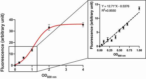
Figure 2. Confocal microscopy Z-stack imaging to visualize C. albicans adhering to saliva-coated beads. The C. albicans SC5314 cells that attached to hydroxyapatite beads were stained with NR. Confocal images were captured with a Zeiss LSCM780 laser scanning microscope. Scale-bar: 20 µm. (A) Bead that had been incubated with C. albicans SC5314 cells at OD600nm = 1, (B) Bead that had been incubated with C. albicans SC5314 cells at OD600nm = 0.1
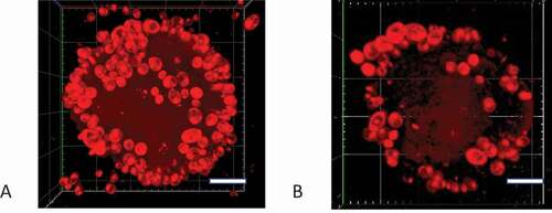
Figure 3. Susceptibility of C. albicans strains deleted in BGL2 and ECM33 to Calcofluor white (CFW). (A) C. albicans SC5314 (wild-type), bgl2∆/∆ (BGL2M4), ecm33∆/∆ (ECM33M4), and bgl2∆/∆,ecm33∆/∆ (BEM4) were serially diluted and spotted onto YPD agar plates and YPD agar plates containing 12.5 μg/mL CFW and incubated at 30°C for 24 h; (B) C. albicans SC5314 (wild-type), and restored strains bgl2Δ/BGL2 (BGL2MK2), ecm33Δ/ECM33 (ECM33MK2), and bgl2Δ/BGL2,ecm33Δ/ECM33 (BEMK2) were serially diluted and spotted onto YPD agar plates and YPD agar plates containing 12.5 μg/mL CFW and incubated at 30°C for 24 h: (C) C. albicans SC5314 (wild-type), als1∆/∆ (ALS1M4), als1∆/∆,bgl2∆/∆ (ABM4), als1∆/∆,ecm33∆/∆ (AEM4), and als1∆/∆, bgl2∆/∆, ecm33∆/∆ (ABEM4) were serially diluted and spotted onto YPD agar plates and YPD agar plates containing 12.5 μg/mL CFW and incubated at 30°C for 24 h
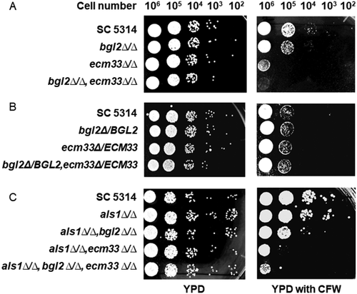
Figure 4. Adherence of C. albicans SC5314 cells to HA beads coated with various proteins. C. albicans cells (1 x 10⁷) were incubated with uncoated, saliva-, skim milk (SM)-, fetal bovine serum (FBS)-, or bovine serum albumin (BSA)-coated HA beads (12 mg) at 28°C for 1 h. The percentage of the input C. albicans cells attached to the HA beads was calculated. Results are the means (± SD) of at least three independent experiments. Asterisks indicate significant differences to the adherence to uncoated HA beads (*** p < 0.001, **** p < 0.0001)
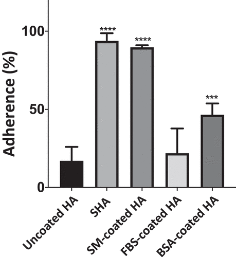
Figure 5. Adherence of wild-type C. albicans SC5314 cells and homozygous BGL2, ECM33, and ALS1 single, double, and triple mutants to SHA beads. Wild-type or mutant C. albicans cells (1 x 10⁷) were incubated with SHA beads (12 mg) at 28°C for 1 h. The results are presented as percentage adherence relative to SC5314, which is set to 100%. Results are the means of at least three independent experiments (± SD). Asterisks indicate significant differences to the adherence of SC5314 cells (*** p < 0.001, **** p < 0.0001)
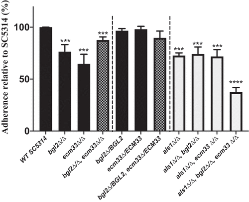
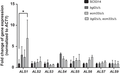
Figure 6. Expression of C. albicans ALS genes in wild-type and mutant strains. C. albicans strains were cultured in YPD medium and then total RNA was isolated and subjected to qRT-PCR. Gene expression was normalized against the C. albicans ACT1 gene and is reported for each ALS gene relative to expression in wild-type strain SC5314. All experiments consisted of triplicate measurements (technical replicates) and experiments were carried out three times (biological replicates) (± SD). (*p < 0.05)