Figures & data
Table 1. Overview of bacterial species and strains used in this study
Figure 1. Effects of repeated rinsing with potential prebiotic substrates at 1 M on multi-species biofilm composition
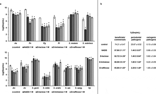
Figure 2. Effects of repeated rinsing with potential prebiotic substrates at 1%(w/v) on multi-species biofilm composition
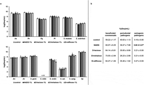
Figure 3. Effects of repeated rinsing with potential prebiotic substrates on multi-species biofilm organic acid balances
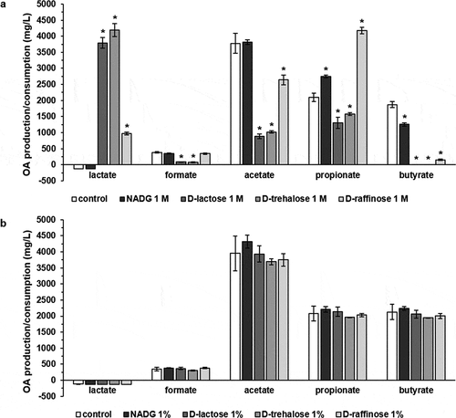
Table 2. Effects of repeated rinsing of multi-species biofilms with potential prebiotic substrates on virulence gene expression from A. actinomycetemcomitans.
Table 3. Effects of repeated rinsing of multi-species biofilms with potential prebiotic substrates on virulence gene expression from P. gingivalis.
Table 4. Effects of repeated rinsing of multi-species biofilms with potential prebiotic substrates on virulence gene expression from F. nucleatum.
Table 5. Effects of repeated rinsing of multi-species biofilms with potential prebiotic substrates on virulence gene expression from P. intermedia.
Table 6. Effects of repeated rinsing with potential prebiotic substrates on multi-species biofilm inflammatory potential towards human oral keratinocytes
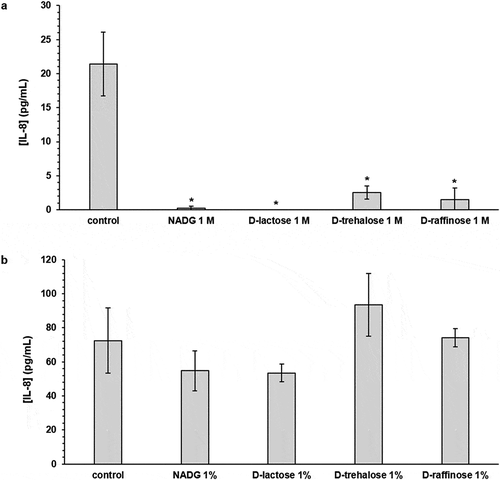
Figure 4. Effects of repeated rinsing with potential prebiotic substrates on multi-species biofilm inflammatory potential