Figures & data
Table 1. Predisposing host factors associated with oral candidiasis and mucormycosis
Table 2. Investigations and management performed for mucormycosis and oral candidiasis
Figure 1. Gram stain shows budding yeast like cells with pseudo hyphae along with few epithelial cells, no pus cells, numerous Gram-negative rods and Gram-positive cocci in pairs
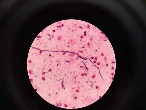
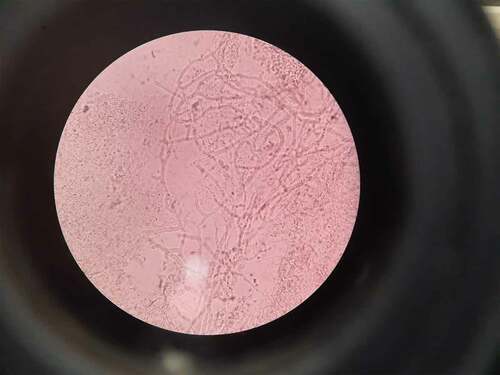
Figure 2. Aseptate hyaline broad branching hyphae on KOH wet mount smear for mucormycosis; site-nasal cavity