Figures & data
Figure 1. NIM screenshot figure of 400% improvement of motor evoked potential in the 6-minute interval for ND of posterior tibial and plantar nerves.
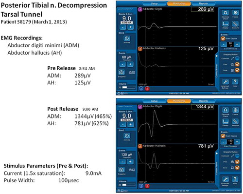
Figure 2. Indentation of common peroneal nerve at R fibular neck is noted just after decompression by division of peroneus longus fascia and a few muscle fibers. Magnification = 1.5×. Patella at 12 o’clock direction, foot to 4:30. With permission of Dr. S. Barrett, Phoenix, AZ.
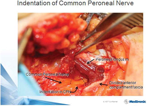
Figure 3. A Kaplan Meier survival curve illustrates the ulcer-free survival of 42 cases with prior healed unilateral DFU and subsequent ND of that leg only. The previously intact contralateral leg, without ND, has a relative risk of subsequent ulceration of 5.5 (p = 0.048). From Nickerson and Rader, JAPMA 104:66–70 (2014), with permission.