
Figures & data
Table 1. Features of the two study groups: non-debrided patients (NDP) and debrided patients (DP).
Figure 1. Steps in the treatment of the diabetic foot by PDT. (a) Molecular structure of the photosensitizers methylene blue and toluidine blue. A (1:1) mixture of two aqueous solutions containing each 1% (mass:mass) of the dyes was used in the treatments. (b) The photosensitizer solutions were applied topically and also injected in the ulcers. (c) A broadband emitter (400–725 nm, maximum at 560 nm) containing a white halogen light, was connected to optical fibers (1.0 or 1.5 mm), which were covered with clear PVC films and introduced in the ulcer cavities to allow efficient illumination of the infected sites. (d) Irradiation was performed weekly with a dose of 6 J/cm2. (e) Subsequently, a LED array with 590 nm and 640 nm centered LEDs was used to illuminate the surface of the infected sites. (f) Irradiations were performed either in close proximity to the lesion or 3–5 cm away from the feet always with a dose of 30 J/cm2. For further details please refer to the Methods section.
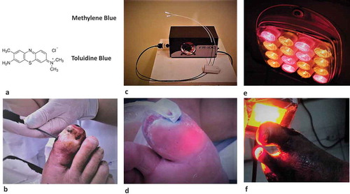
Table 2. Statistical comparison between DP and NDP groups before PDT treatment.
Figure 2. Images of patients before and after PDT. (a, b) Patient from the NDP group, was treated for 62 days to complete healing (28/04–29/06). (c, d) Patient from the DP group, had his lesion healed in 125 days (08/12–14/04).
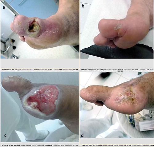
Figure 3. Osteomyelitis before and after PDT. X-Ray images obtained from patients with osteomyelitis before and after PDT treatment. A, B, C, and D are X-ray images from four patients treated with PDT. Arrows at left are showing osteomyelitis in metatarsal and phalanges before treatment with cortical disruption and pathological destruction or disappearance of bone tissue. Arrows at right represent the same patients after PDT. Note the effective regeneration of diseased bones after PDT. A and B were obtained from patients of the DP group, C and D from the NDP group.
Table 3. Statistical comparison between DP and NDP groups after PDT treatment.