Figures & data
Figure 1. Nucleotide sequence analysis showing a germline two base monoallelic deletion in CYLD exon 11 in two brothers presenting with multiple cutaneous cylindromas on the scalp (white arrowheads). The asterisk indicates a STOP codon predicted to terminate translation. Nucleotides deleted in the wild-type allele are underlined.

Figure 2. Pedigree of the four-generation family with the Brooke–Spiegler (CYLD) skin tumor syndrome.
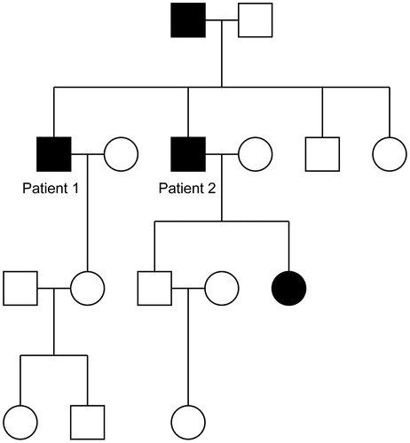
Table 1. CYLD primers used for PCR amplification and nucleotide sequencing.
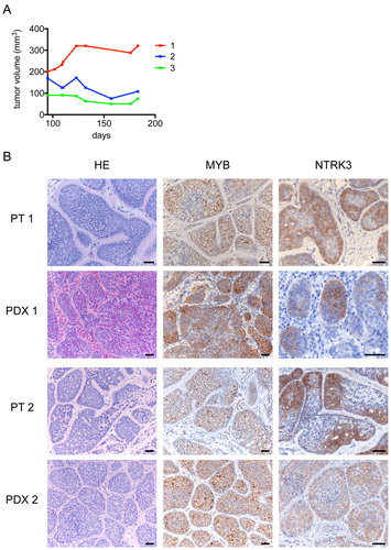
Figure 3. Growth characteristics and morphology of patient-derived primary cylindroma xenografts (PDXs). (A) Growth curves of three cylindroma PDXs. (B) Hematoxylin and Eosin (HE), MYB and NTRK3 immunostainings in primary cylindromas and PDX tumors. PT 1: primary tumor 1; PDX 1: PDX from primary tumor 1; PT 2: primary tumor 2; PDX 2: PDX from primary tumor 2; PDX 3 (panel A): PDX from primary tumor 2. Scale bars indicate 50 µm.