
Figures & data
Figure 1. Initial CR of a 59 year old woman with a comminute intra-articular distal radial fracture and a fracture of the ulnar styloid process. (a) AP view. (b) Sagittal view showing dorsal tilting of the radial articular surface.
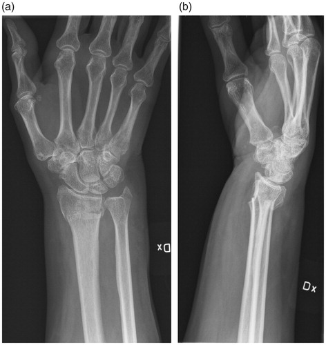
Figure 2. Preoperative CR of the same patient as in Figure 1. One year later, during conservative treatment, the distal radial fracture has healed in malunion and the patient is scheduled for corrective osteotomy. (a) AP view showing axial shortening. (b) Sagittal view showing severe volar tilt.
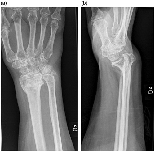
Figure 3. First postoperative control of the same patient as in Figures 1 and 2.The open-wedge corrective osteotomy is fixated with a volar plate. (a) AP view showing axial shortening and ulnar position is now restored. (b) Sagittal view showing volar tilt close to normal.
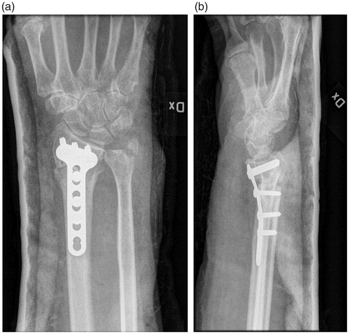
Figure 4. Long-term follow-up, 9 years post-operatively, of the same patient as in Figures 1–3. (a) AP view showing development of a slight axial shortening. No osteoarthritis is seen. (b) Sagittal view showing that the major improvement in joint positioning is maintained.
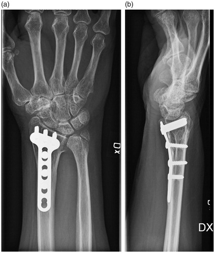
Table 1. Demographics.
Table 2. Number of patients related to the level of osteoarthritis pre-operatively (N = 37) and at follow-up (N = 36), measured using the scale of Knirk and Jupiter.
Table 3. Grip strength (KG) and range of motion (degrees).
Table 4. RAND-36 scores N = 37 median (min–max), mean (SD).