
Figures & data
Figure 1. The incision in a cadaver. The previous incision is extended proximally and the carpal tunnel is opened. *: median nerve; Black arrow: ulnar leaf of flexor retinaculum; White arrow: palmaris longus tendon.
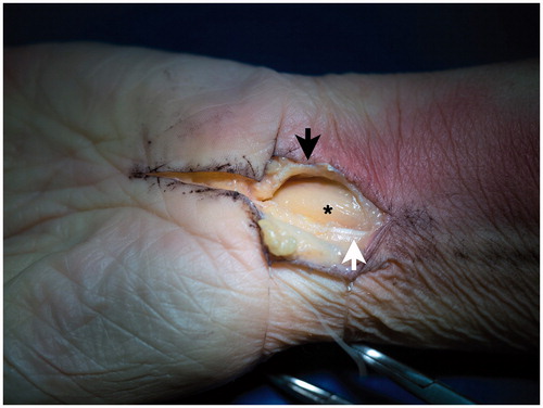
Figure 2. Intraoperative appearance in a cadaver. The palmaris longus tendon is dissected proximally. *: median nerve; Black arrows: flexor retinaculum; White arrow: palmaris longus tendon.
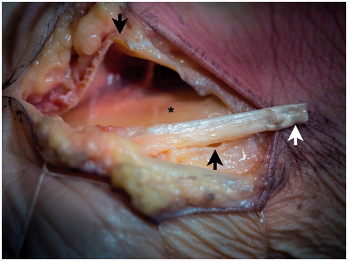
Figure 3. Interposition of the palmaris longus tendon in a cadaver. The palmaris longus tendon is flipped distally and sutured between the edges of the flexor retinaculum. *: median nerve; Black arrows: flexor retinaculum; White arrow: palmaris longus tendon.
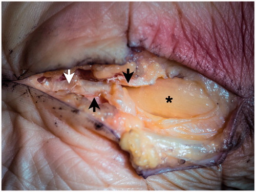
Table 1. Patient characteristics of the study sample (n = 20 hands).
Table 2. Pre- and intraoperative findings and postoperative outcomes.