
Figures & data
Figure 1. Examples of biopsies. Masson trichrome staining, original magnification 40x (a,d); 100x (b,e); α-smooth muscle actin, original magnification 40x (c,f). (A–C) illustrates a type A reaction and d-f a type B reaction. Small arrow: synovial metaplasia. Arrow head: mesh fibers. Asterix: collagen fibers. Thick arrow: actine positive myofibroblasts.
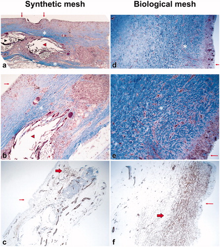
Table 1. Morphological evaluation of the biopsies.
Table 2. Result of actin immunohistostaining.