
Figures & data
Figure 1. (A) Schematic view of the forearm three segments division. The circles in red highlights the proximal third which is the object of investigation in our anatomical study. P: pisiform bone; ME: medial epicondyle. (B) A volar side, skin incision, parallel of the longitudinal axis of the forearm, was performed in the proximal third. A supra fascial dissection in radio-ulnar direction under 4.0× loupes magnification was conducted to visualize the perforators. FRC: flexor carpi radialis muscle; FUC: flexor carpi ulnaris muscle; ME: medial epicondyle; *: perforator branch. Scale bar (1 cm).
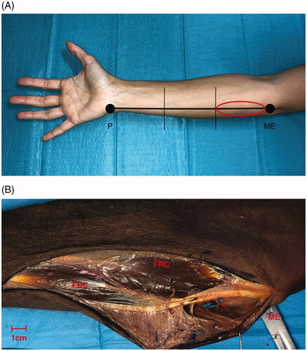
Figure 2. Anatomy of the upper limb in the proximal third (volar side) focusing on the ulnar artery (UA). Overturning proximally the insertion of the flexor carpi ulnaris muscle (FUC), the first tract of the UA is exposed running on the flexor digitorum profundus muscle (FDP): it gives firstly the recurrent branches on the ulnar edge, following by the common ulnar interosseous division on the radial side. Secondly, between its the proximal and middle third the UA gives a further branch on the ulnar side (**). Legend: (*) Anterior recurrent ulnar perforator artery, (**) direct ulnar perforator artery, cutaneous end point (CE), flexor carpi ulnaris muscle (FUC) and flexor digitorum profundus muscle (FDP). Scale bar (1 cm).
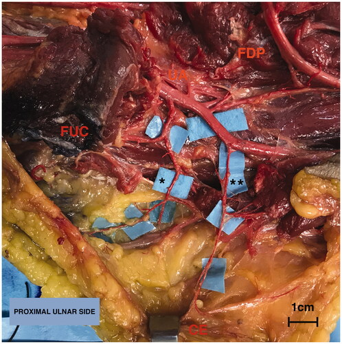
Figure 3. Single case (one out eight specimens) in which the perforator (*) originated directly from the brachial artery above the ulnar fold. The red string indicates the brachial artery (BA). Flexor carpi ulnaris muscle (FUC). Scale bar (1 cm).
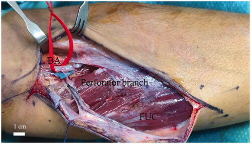
Table 1. Number of perforators, diameters at the origin and at the perforator point of the ulnar perforator or anterior recurrent ulnar perforator artery in the eight injected specimens dissected.
Table 2. Distances between the origin and medial humeral epicondyle or distances between the cutaneous end point and medial humeral epicondyle and the pedicle length for each type of perforator (direct ulnar perforator or anterior recurrent ulnar perforator) in the eight injected specimens dissected.