
Figures & data
Figure 1. Mechanistic model of CVD risk in PTSD.
Note. BP = blood pressure; HR = heart rate; HRV = heart rate variability; NTS = solitary nucleus; RVLM = rostral ventrolateral medulla; SNA = stress-associated neural network activity; vmPFC = ventromedial prefrontal cortex. PTSD leads to alterations in stress-related neural activity, leading to increased inflammation and autonomic dysfunction, which both lead to cardiovascular disease.
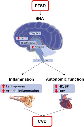
Figure 2. Study design overview.
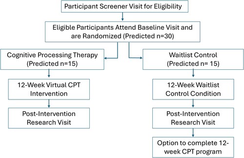
Table 1. Schedule of study assessments.
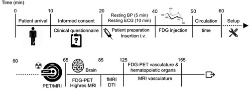
Figure 3. Overview of imaging session.
Note. BP = Blood Pressure; ECG = electrocardiogram; FDG = 18F-fluorodeoxyglucose; PET/MRI = positron emission tomography/ magnetic resonance imaging; fMRI = functional magnetic resonance imaging; DTI = diffusion tensor imaging; all neural imaging is performed at rest besides fMRI which is performed with eyes opened with fixation cross and the Hariri task. Timeline of procedures for the 155-minute imaging session. The first 60 minutes of the session includes consent, measure of autonomic function, participant preparation and set-up followed by FDG-PET/MRI brain and peripheral imaging.
Data availability statement
Data sharing not applicable – no new data generated.