
Figures & data
Figure 1. Oculomotor network. Visuospatial sensory information obtained from oculomotion travels to the superior colliculus in the midbrain via cranial nerves III, IV, and VI. The superior colliculus can project visuospatial afferents to the frontal eye field (FEF) to engage the dorsal visual stream, which is a functional component of the dorsal attentional network that helps guide one’s visuospatial processing of the external environment. The FEF functionally connects with the lateral posterior parietal cortex (Par), where one can process visuospatial details related to one’s viewer-centred egocentric perspective (i.e. identifying one’s self-location). The FEF also interacts with the supplementary eye field (SEF), which maintains connections with both the lateral and medial parietal cortices. The SEF, through its connections with the parietal cortex, can process visuospatial details from both an egocentric and observer-centred allocentric perspective, as it can identify one’s self-location based on identifying objects or external locations in the environment. The eye clipart image was retrieved and adapted from a free public domain (clker.com, Rolera LLC).
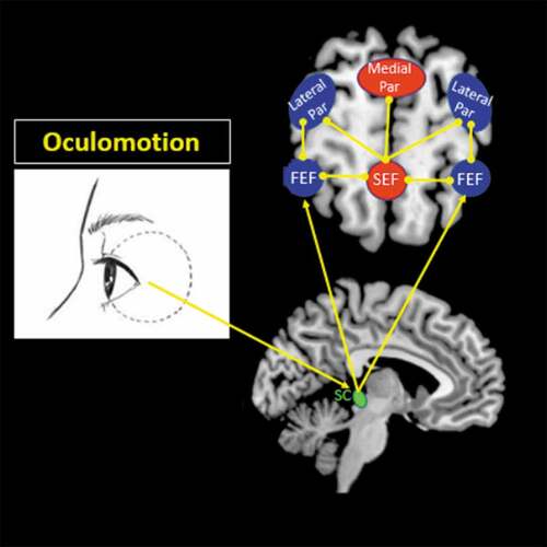
Table 1. Clinical and demographic information.
Figure 2. Experimental paradigm. All participants were asked to retrieve both neutral and traumatic autobiographical memories via a personalized word cue associated with each memory, while following a moving dot to guide eye movements across the screen. In total, there were three conditions, each lasting 13 minutes, conducted in the following order: no memory retrieval, neutral memory retrieval, and traumatic memory retrieval. Each condition consisted of 12 runs, separated into four blocks to present each type of oculomotor stimulus in three consecutive runs (stationary fixation dot, a horizontal smooth pursuit, a horizontal saccadic pursuit, and a vertical saccadic pursuit). Each run lasted (6 + 3 + 30) 39 seconds, where a black central stationary dot was displayed for 6 seconds to obtain an implicit baseline measure, after which participants were instructed to retrieve autobiographical memories while reading a single personalized word cue displayed on the screen for 3 seconds (replaced with a ‘+’ symbol in the no memory retrieval condition). Immediately afterwards, participants were asked to continue retrieving the memory while 30 seconds of one type of oculomotor stimulus was presented using coloured circles to guide eye movements across the screen. After three consecutive runs using the same type of oculomotor stimulus, participants were asked to rate the severity of post-traumatic stress disorder (PTSD) symptoms they experienced during memory retrieval with the specific adjunctive oculomotor stimulus, including emotional intensity, numbing, dissociation, re-experiencing, and vividness of memory. Afterwards, an 18-second rest interval using a black stationary fixation ‘+’ led to a transition into a new type of oculomotion. This process was repeated four times to evaluate the effects of each type of oculomotor stimulus.
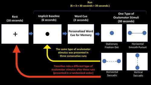
Table 2. Right frontal (FEF) and supplementary eye field (SEF) psychophysiological interaction post-hoc two-sample t-tests and correlations with clinical dissociative symptoms.
Figure 3. Explorative functional connectivity analyses (psychophysiological interaction) of (A) the right frontal eye field [FEF (x: 46, y: 0, z: 56)] and (B) the right supplementary eye field [SEF (x: 2, y: 2, z: 62)] seed regions during the traumatic memory retrieval condition. (A) During retrieval of a traumatic/stressful memory, as compared to the post-traumatic stress disorder (PTSD) patient group, healthy controls demonstrated increased right FEF connectivity with the right posterior insula with simultaneous smooth pursuit eye movements. In contrast, as compared to healthy controls, PTSD patients demonstrated increased right FEF connectivity with the right dorsolateral prefrontal cortex during retrieval of a traumatic memory with simultaneous smooth pursuit eye movements. (B) During retrieval of a traumatic/stressful memory with smooth pursuit eye movements, as compared to healthy controls, PTSD patients showed increased right SEF connectivity with the right dorsomedial and the right dorsolateral prefrontal cortices. In addition, as compared to controls, PTSD patients showed increased right SEF connectivity with the right anterior insula during retrieval of a traumatic memory with concurrent saccadic eye movements. All results are shown at pFWE ≤ 0.0125, k = 10, to correct for multiple comparisons; however, the precuneus is pFWE whole-brain corrected at p < 0.05, k = 10.
![Figure 3. Explorative functional connectivity analyses (psychophysiological interaction) of (A) the right frontal eye field [FEF (x: 46, y: 0, z: 56)] and (B) the right supplementary eye field [SEF (x: 2, y: 2, z: 62)] seed regions during the traumatic memory retrieval condition. (A) During retrieval of a traumatic/stressful memory, as compared to the post-traumatic stress disorder (PTSD) patient group, healthy controls demonstrated increased right FEF connectivity with the right posterior insula with simultaneous smooth pursuit eye movements. In contrast, as compared to healthy controls, PTSD patients demonstrated increased right FEF connectivity with the right dorsolateral prefrontal cortex during retrieval of a traumatic memory with simultaneous smooth pursuit eye movements. (B) During retrieval of a traumatic/stressful memory with smooth pursuit eye movements, as compared to healthy controls, PTSD patients showed increased right SEF connectivity with the right dorsomedial and the right dorsolateral prefrontal cortices. In addition, as compared to controls, PTSD patients showed increased right SEF connectivity with the right anterior insula during retrieval of a traumatic memory with concurrent saccadic eye movements. All results are shown at pFWE ≤ 0.0125, k = 10, to correct for multiple comparisons; however, the precuneus is pFWE whole-brain corrected at p < 0.05, k = 10.](/cms/asset/321e8a5c-4fa6-4677-abc4-3011f35ea171/zept_a_1586265_f0003_oc.jpg)
Figure 4. Explorative negative functional connectivity correlations with clinical dissociative measures in the right supplementary eye field (SEF) psychophysiological interaction during the traumatic memory retrieval condition. During retrieval of a traumatic memory with horizontal smooth pursuit eye movements, trait dissociation [Multiscale Dissociation Inventory (MDI)] symptoms and state dissociation symptoms [Responses to Script-Driven Imagery Scale (RSDI)] measures collected just prior to the scan correlated negatively with right SEF connectivity with the right dorsolateral prefrontal cortex. Pos, posterior; Ant, anterior. Results are shown at pFWE ≤ 0.0125, k = 10, corrected for multiple comparisons.
![Figure 4. Explorative negative functional connectivity correlations with clinical dissociative measures in the right supplementary eye field (SEF) psychophysiological interaction during the traumatic memory retrieval condition. During retrieval of a traumatic memory with horizontal smooth pursuit eye movements, trait dissociation [Multiscale Dissociation Inventory (MDI)] symptoms and state dissociation symptoms [Responses to Script-Driven Imagery Scale (RSDI)] measures collected just prior to the scan correlated negatively with right SEF connectivity with the right dorsolateral prefrontal cortex. Pos, posterior; Ant, anterior. Results are shown at pFWE ≤ 0.0125, k = 10, corrected for multiple comparisons.](/cms/asset/336149bf-b987-4a8c-a5e0-fba085d7aee1/zept_a_1586265_f0004_oc.jpg)