Figures & data
Figure 1. R. africae infected Vero-cells showing rickettsial organisms stained red with Gimenez (a) and green with immunofluorescent anti-rickettsial goat anti-rabbit immunoglobulin (b). Original magnification × 1000.
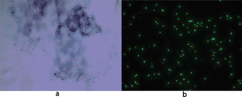
Table 1. Summary of PCR results and immunofluorescent titres of acute and convalescent sera from Patients 1 and 2 using R. helvetica and R. africae as antigen, respectively.
Figure 2. Western blot analysis of IgG antibodies against R. africae whole cell antigen. Lane P2 demonstrates the lipopolysaccharide ladders (LPS) and specific reactions against spotted fever rickettsia proteins in the 110–150 kDa region for convalescent serum for Patient 2.