
Figures & data
Table 1. experimental design of bacteriophage treatments in (SPF) chicks challenged with S. typhimurium and S. enteritidis.
Table 2. Oligonucleotide primers and probes used in this study.
Figure 1. Plaque assay for bacteriophage enumeration.
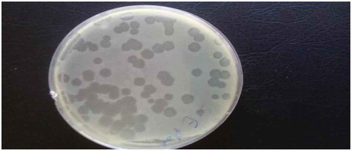
Figure 2. (a) hemorrhagic patches in liver with distention of intestine with diarrhea (b) congestion of internal organs with pasting vent (c) enlargement and distention of the two ceci with diarrhea.
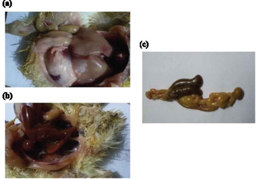
Table 3. Quantitative detection of S. typhimurium colonization in cecum of chicks under experiment.
Table 4. Quantitative detection of S. enteritidis colonization in cecum of chicks under experiment.