Figures & data
Table 1. Cases of esophageal pneumatosis reported.
Figure 1. Computed tomogram of the abdomen shows gastric pneumatosis and hepatic portal venous gas extending the entire venous tree and to the liver margins. Intramural gas in the stomach.
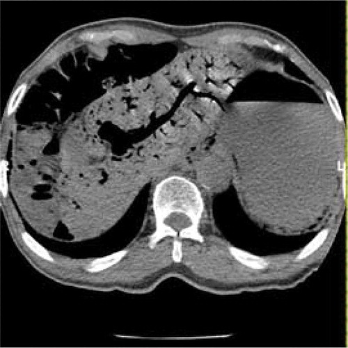
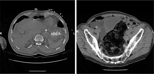
Figure 2. Computed tomogram of the abdomen revealing intramural gas in the stomach extending to the visualized esophagus. The small bowel is diffusely gas distended, but there is no intramural gas noted.
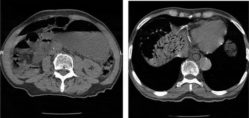
Figure 3. Computed tomogram of the abdomen showing resolution of gastric pneumatosis as well as abundant stool in the rectum.