Figures & data
Figure 1. Axial computed tomography showing a low attenuating right adrenal mass, which proved to be an adrenal collision tumor on pathology.
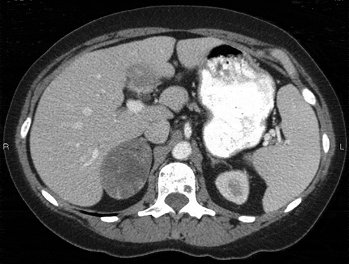
Figure 2. Coronal computed tomography showing a low attenuating right adrenal mass, which proved to be an adrenal collision tumor on pathology.
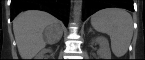
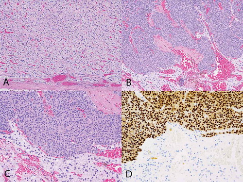
Figure 3. Histological findings of the patient. (A) Hematoxylin & eosin stain showing adrenal cortical adenoma with a capsule of adenoma noted at the bottom of the image (100×). (B) Metastatic breast carcinoma (top of image) and adrenal cortical adenoma (bottom) (100×). (C) Metastatic breast carcinoma (top of image) and adrenal cortical adenoma (bottom) (200×). (D) GATA-3 immunohistochemical stain showing metastatic breast carcinoma (top of image) and adrenal cortical adenoma (bottom). The metastatic breast carcinoma shows positive nuclear staining (brown) with the GATA-3 antibody consistent with breast carcinoma, while the cells of the adrenal cortical adenoma are negative for that stain (200×).