Figures & data
Figure 1. Left: H&E stain of left breast biopsy. Right: Left breast mass biopsy staining for Her2.
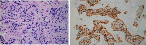
Figure 2. Left: MRI image of solitary 3.5cm mass in uncinate process of pancreas (arrow). Right: MRI image of diffusely thickened stomach wall (arrow).
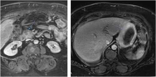
Figure 3. EGD visualizing abnormal lesions in stomach demonstrating areas concerning for malignancy (arrows).
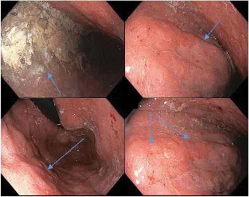
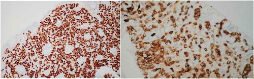
Figure 4. Left: stomach lesion biopsy staining for GATA3. Right: pancreatic lesion biopsy staining for E-Cadherin.