899
Views
3
CrossRef citations to date
0
Altmetric
Case Report

An atypical subacute presentation of posterior reversible encephalopathy syndrome

ORCID Icon, &
Pages 269-274 | Received 11 May 2017, Accepted 07 Aug 2017, Published online: 19 Sep 2017

Figures & data

Figure 1. Fluid-attenuated inversion recovery (FLAIR) magnetic resonance images (MRI) at presentation showed scattered regions of hyperintense signal in the frontal, parietal, occipital, and temporal lobes bilaterally (a–d). FLAIR hyperintensities are also seen in the deep grey nuclei (b), midbrain (d–e), and cerebellum (e–f).
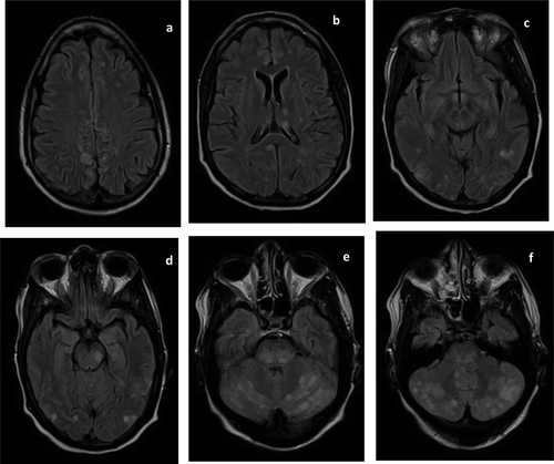
Figure 1. Fluid-attenuated inversion recovery (FLAIR) magnetic resonance images (MRI) at presentation showed scattered regions of hyperintense signal in the frontal, parietal, occipital, and temporal lobes bilaterally (a–d). FLAIR hyperintensities are also seen in the deep grey nuclei (b), midbrain (d–e), and cerebellum (e–f).

Figure 2. At presentation, magnetic resonance angiogram (MRA) of the circle of Willis showed no abnormality.
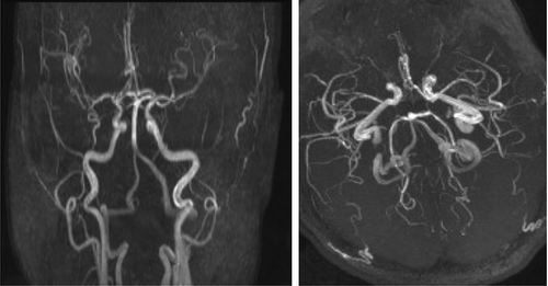
Figure 2. At presentation, magnetic resonance angiogram (MRA) of the circle of Willis showed no abnormality.

Figure 3. Repeat MRI done on hospital day 7 showed resolution of the previously noted extensive signal abnormalities. However, two punctate foci of FLAIR hyperintensities persisted in the right corona radiata (b) and left frontal lobe (c). These may represent age-related white matter changes or white matter hyperintensities of presumed vascular origin.
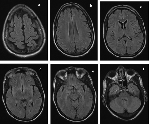
Figure 3. Repeat MRI done on hospital day 7 showed resolution of the previously noted extensive signal abnormalities. However, two punctate foci of FLAIR hyperintensities persisted in the right corona radiata (b) and left frontal lobe (c). These may represent age-related white matter changes or white matter hyperintensities of presumed vascular origin.