Figures & data
Figure 1. CT of abdomen and pelvis with intravenous contrast revealing right-sided PA with multiloculation and contiguous spread, as shown by the arrows in the coronal (left) and sagittal (right) sections.
![Figure 1. CT of abdomen and pelvis with intravenous contrast revealing right-sided PA with multiloculation and contiguous spread, as shown by the arrows in the coronal (left) and sagittal (right) sections.](/cms/asset/f968d4f9-2f52-42ad-a2fc-12509f416177/zjch_a_1433431_f0001_oc.jpg)
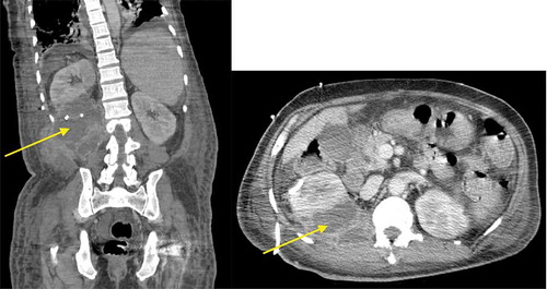
Table 1. Case reports of GBS intra-abdominal abscesses.