Figures & data
Figure 2. Purpura involving the ear.
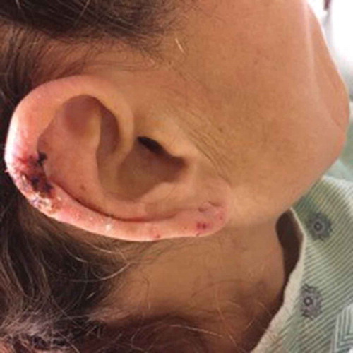
Figure 3. Retiform purpura in the leg.
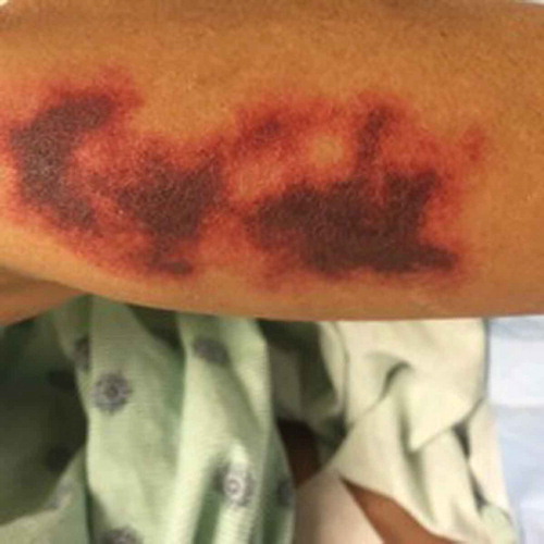
Figure 4. 2X. Sections show a punch biopsy of skin. There is subepidermal edema with erythrocyte extravasation, Many superficial and deep dermal small vascular channels are surrounded by neutrophils and nuclear dust.
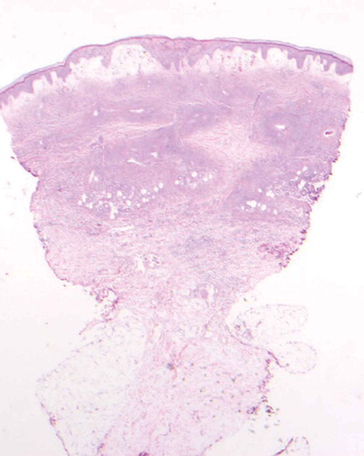
Figure 5. 40X. Neutrophils and nuclear dust in the blood vessel walls causing destruction of the blood vessel walls.
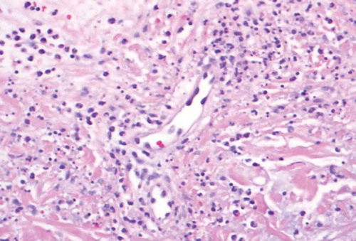
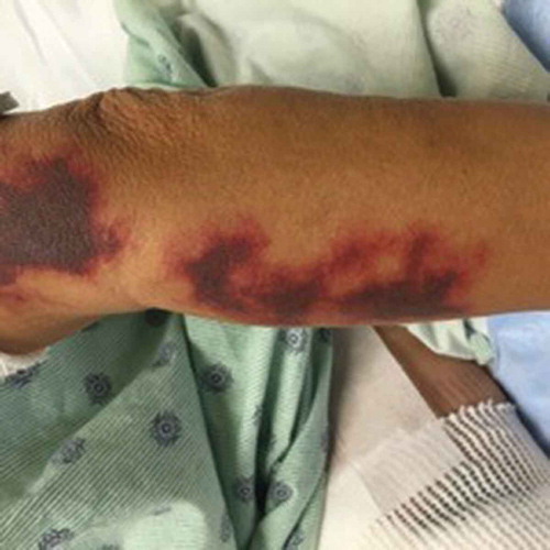
Figure 1. Retiform purpura in the arms.