Figures & data
Figure 1. CT scan.
Bowel wall thickening at the level of distal descending and sigmoid colon indicated by a red arrow – CT scan.
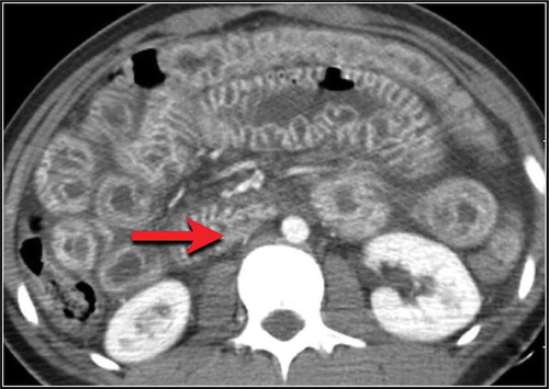
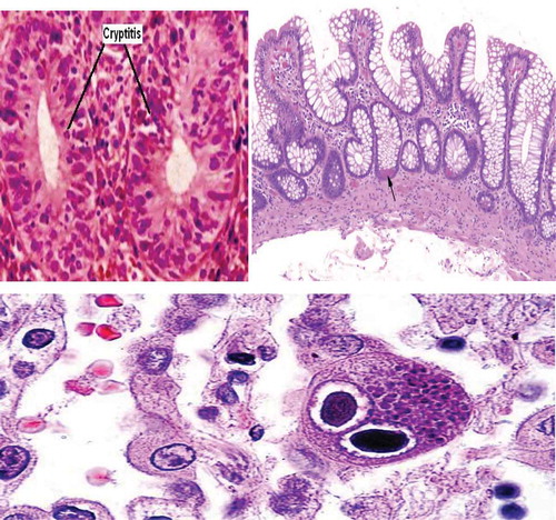
Figure 2. Colonoscopic biopsy.
On above Left Image: Hematoxylin and eosin staining showing Cryptitis. On above right side: Hematoxylin and Eosin staining showing shortening of the crypts and Crypts Abscess – marked by Black Arrow. On below image: classic ‘owl eye inclusion bodies’ visualized on histology
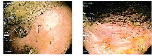
Colonoscopy image