Figures & data
Figure 1. CT chest showing bilateral lobar pneumonia.
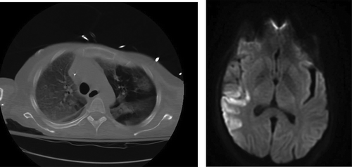
Figure 2. MRI brain showing acute ischemic edema involving the right posterior frontal parietal distribution.
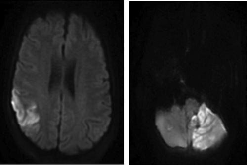
Figure 3. MRI brain showing acute ischemic edema in the left cerebellar hemisphere.
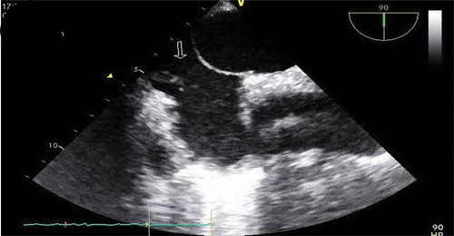
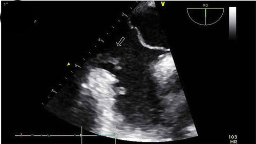
Figure 4. TEE showing vegetations on Eustachian valve (arrows).