
Figures & data
Figure 1. Entake PEG safety system (Commed, Utica, NY). The tube dome is collapsible upon pulling allowing passage of the tube through the gastrocutaneous tract.
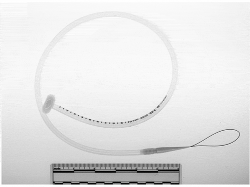
Figure 2. Gastric wall defect 6 hours after inadvertent removal of PEG tube (white arrow). Note partial sealing of the defect.
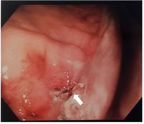
Figure 3. Endoscopic view of double-PEG, placed to improve the apposition and patching of the gastric and abdominal walls.
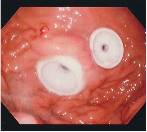
Table 1. Clinical data (*indicates time from attempted direct tube replacement [DTR]).