Figures & data
Figure 1. A transesophageal echocardiogram showing a large echogenic mass (blue arrow) measuring 14 mm x 7 mm on ventricular aspect in outflow tract.
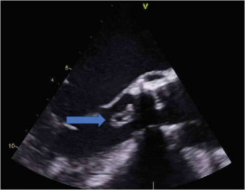
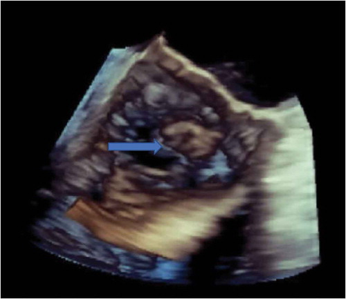
Figure 2. 3-D Echocardiogram identifying vegetation (blue arrow) on the non-coronary cusp of aortic valve.
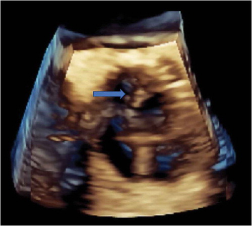
Figure 3. 3-D Echocardiogram showing vegetation (blue arrow) on the non-coronary cusp of aortic valve.