Figures & data
Figure 1. (a) CXR demonstrating pulmonary vascular markings with increased cephalization. (b) Electrocardiogram demonstrating normal sinus rhythm and no signs of ST-elevation or depression, no inverted T waves noted.
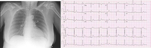
Figure 2. a–d: (a, b) Cine shot of coronary vasculature showing no evidence of obstructive coronary disease. (c) Left ventriculogram push demonstrating focal wall hypokinesis. (d) Transthoracic echocardiogram demonstrating EF: 40%.
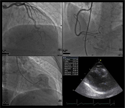
Figure 3. Left ventriculogram showing reverse ballooning during systole.
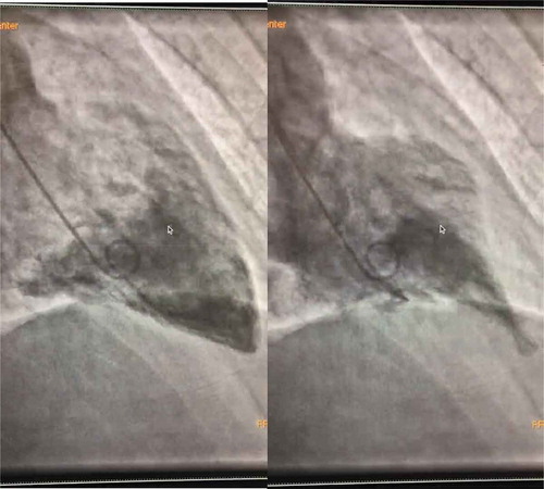
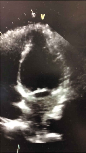
Figure 4. Echocardiogram also revealing basal ballooning in this reverse variant of Takotsubo cardiomyopathy.