
Figures & data
Table 1. Workup of to evaluate secondary causes of hypertension in our patient.
Figure 1. MRI abdomen/pelvis without contrast demonstrating bilateral solid slightly heterogeneous adrenal gland masses (yellow arrows).
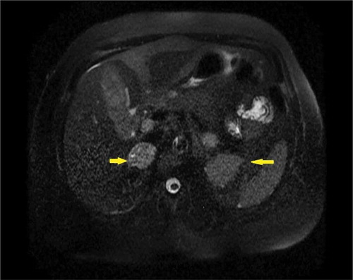
Figure 2. PET scan showing intense FDG (fluorodeoxyglucose) localization within the enlarged right adrenal gland (yellow arrow).
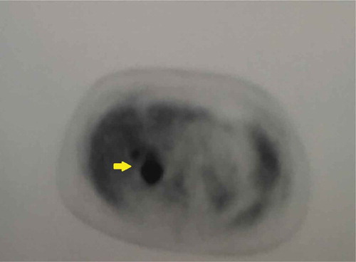
Table 2. National institutes of health 1988 consensus development conference diagnostic criteria for Neurofibromatosis −1.
Table 3. Conditions associated with Neurofibromatosis-1.