Figures & data
Figure 1. Positive ice pack test. A marked improvement of ptosis is noted after ice pack application for 2 minutes.
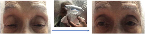
Figure 2. Bone marrow biopsy slides, demonstrating near absence of erythroid elements. (a) Low-power view of a HE-stained section (x20). (b) High-power view of a HE-stained section (x400). (c) Low-power view of a CD71 stain for confirmation of pure red cell aplasia.
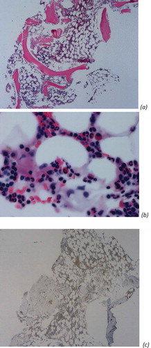
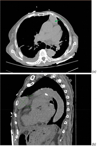
Figure 3. Computed tomography (CT) chest without contrast revealed ovoid anterior mediastinal mass consistent with thymoma. (a) Green arrow. (b) Diameter measurement of thymoma, 5.6 × 3.1 cm ovoid anterior mediastinal mass.