Figures & data
Figure 1. MRI brain T1 sequence with contrast. Centered in the right Meckel’s cave is a homogeneously enhancing 18 × 11 x 8 mm mass with enhancement coursing along the V2 and V3 segments of the right 5th cranial nerve
Figure 2. Smear preparation showing cohesive clusters of epithelioid cells, scattered background small lymphocytes (Papanicolaou stain, 200x original magnification)
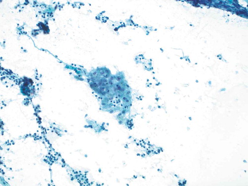
Figure 3. Smear preparation showing cohesive clusters of epithelioid cells, scattered background small lymphocytes (Papanicolaou stain, 200x original magnification)
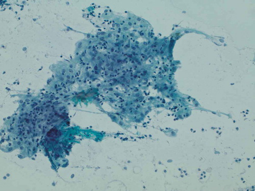
Figure 4. Representative epithelioid cellular cluster showing architectural cohesion, indistinct cytoplasmic borders, and uniform-appearing nuclei with minute nucleoli, consistent with non-caseating granuloma (Papanicolaou stain, 400x original magnification)
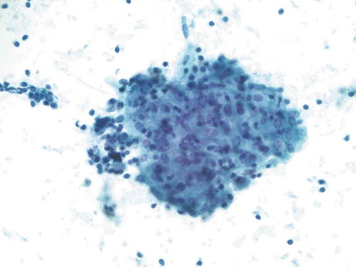
Figure 5. Six-month surveillance imaging: MRI brain T1 sequence with contrast. There is interval decrease in pathologic enhancement involving the right Meckel’s cave mass with new enhancement within left Meckel’s cave