Figures & data
Image 1. EKG: T wave inversion in anterior-lateral and inferior leads
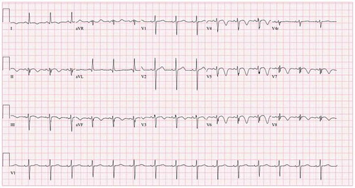
Image 2 ECHO:Non contrast apical four chamber view showing echo density in the left ventricular apex which represents thrombus vs mass
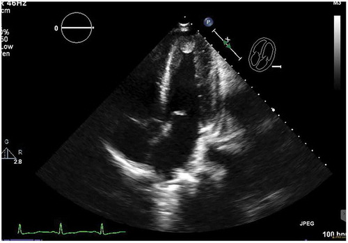
Image 3 ECHO: Image obtained after administration of contrast agent, showing spherical filling defect noted in apex, consistent with apical thrombus
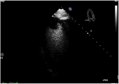
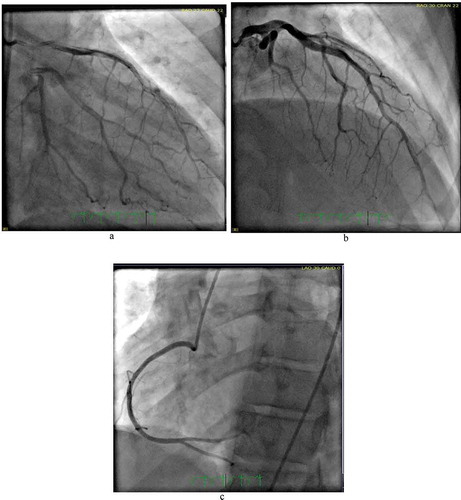
Image 4 Cath: Coronary angiogram showing arterial wall contrast staining with multiple radiolucent lumen in mid to distal left anterior descending artery consistent with type 1 spontaneous coronary artery dissection (SCAD) . Normal right coronary artery